Iron »
PDB 2zph-3a0b »
2zvu »
Iron in PDB 2zvu: Crystal Structure of Rat Heme Oxygenase-1 in Complex with Ferrous Verdoheme
Enzymatic activity of Crystal Structure of Rat Heme Oxygenase-1 in Complex with Ferrous Verdoheme
All present enzymatic activity of Crystal Structure of Rat Heme Oxygenase-1 in Complex with Ferrous Verdoheme:
1.14.99.3;
1.14.99.3;
Protein crystallography data
The structure of Crystal Structure of Rat Heme Oxygenase-1 in Complex with Ferrous Verdoheme, PDB code: 2zvu
was solved by
H.Sato,
M.Sugishima,
K.Fukuyama,
M.Noguchi,
with X-Ray Crystallography technique. A brief refinement statistics is given in the table below:
| Resolution Low / High (Å) | 32.67 / 2.20 |
| Space group | P 32 2 1 |
| Cell size a, b, c (Å), α, β, γ (°) | 65.346, 65.346, 120.240, 90.00, 90.00, 120.00 |
| R / Rfree (%) | 18.9 / 24 |
Iron Binding Sites:
The binding sites of Iron atom in the Crystal Structure of Rat Heme Oxygenase-1 in Complex with Ferrous Verdoheme
(pdb code 2zvu). This binding sites where shown within
5.0 Angstroms radius around Iron atom.
In total only one binding site of Iron was determined in the Crystal Structure of Rat Heme Oxygenase-1 in Complex with Ferrous Verdoheme, PDB code: 2zvu:
In total only one binding site of Iron was determined in the Crystal Structure of Rat Heme Oxygenase-1 in Complex with Ferrous Verdoheme, PDB code: 2zvu:
Iron binding site 1 out of 1 in 2zvu
Go back to
Iron binding site 1 out
of 1 in the Crystal Structure of Rat Heme Oxygenase-1 in Complex with Ferrous Verdoheme

Mono view
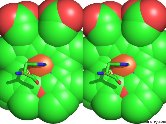
Stereo pair view
Mono view
Stereo pair view
A full contact list of Iron with other atoms in the Fe binding
site number 1 of Crystal Structure of Rat Heme Oxygenase-1 in Complex with Ferrous Verdoheme within 5.0Å range:
|
Reference:
H.Sato,
M.Sugishima,
H.Sakamoto,
Y.Higashimoto,
C.Shimokawa,
K.Fukuyama,
G.Palmer,
M.Noguchi.
Crystal Structure of Rat Haem Oxygenase-1 in Complex with Ferrous Verdohaem: Presence of A Hydrogen-Bond Network on the Distal Side Biochem.J. V. 419 339 2009.
ISSN: ISSN 0264-6021
PubMed: 19154182
DOI: 10.1042/BJ20082279
Page generated: Mon Aug 4 22:57:02 2025
ISSN: ISSN 0264-6021
PubMed: 19154182
DOI: 10.1042/BJ20082279
Last articles
Mn in 9LJUMn in 9LJW
Mn in 9LJS
Mn in 9LJR
Mn in 9LJT
Mn in 9LJV
Mg in 9UA2
Mg in 9R96
Mg in 9VM1
Mg in 9P01