Iron »
PDB 2zph-3a0b »
2zwj »
Iron in PDB 2zwj: Crystal Structure of A Hemoglobin Component V From Propsilocerus Akamusi (PH4.6 Coordinates)
Protein crystallography data
The structure of Crystal Structure of A Hemoglobin Component V From Propsilocerus Akamusi (PH4.6 Coordinates), PDB code: 2zwj
was solved by
T.Kuwada,
T.Hasegawa,
T.Takagi,
F.Shishikura,
with X-Ray Crystallography technique. A brief refinement statistics is given in the table below:
| Resolution Low / High (Å) | 37.63 / 1.81 |
| Space group | P 21 21 2 |
| Cell size a, b, c (Å), α, β, γ (°) | 65.540, 75.260, 33.680, 90.00, 90.00, 90.00 |
| R / Rfree (%) | n/a / n/a |
Iron Binding Sites:
The binding sites of Iron atom in the Crystal Structure of A Hemoglobin Component V From Propsilocerus Akamusi (PH4.6 Coordinates)
(pdb code 2zwj). This binding sites where shown within
5.0 Angstroms radius around Iron atom.
In total only one binding site of Iron was determined in the Crystal Structure of A Hemoglobin Component V From Propsilocerus Akamusi (PH4.6 Coordinates), PDB code: 2zwj:
In total only one binding site of Iron was determined in the Crystal Structure of A Hemoglobin Component V From Propsilocerus Akamusi (PH4.6 Coordinates), PDB code: 2zwj:
Iron binding site 1 out of 1 in 2zwj
Go back to
Iron binding site 1 out
of 1 in the Crystal Structure of A Hemoglobin Component V From Propsilocerus Akamusi (PH4.6 Coordinates)
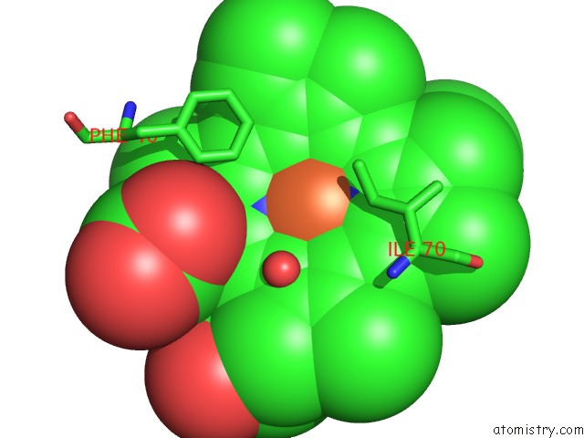
Mono view
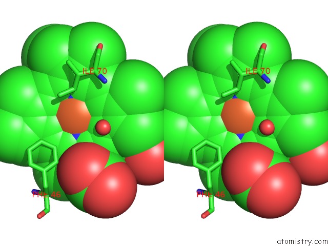
Stereo pair view
Mono view
Stereo pair view
A full contact list of Iron with other atoms in the Fe binding
site number 1 of Crystal Structure of A Hemoglobin Component V From Propsilocerus Akamusi (PH4.6 Coordinates) within 5.0Å range:
|
Reference:
T.Kuwada,
T.Hasegawa,
T.Takagi,
I.Sato,
F.Shishikura.
pH-Dependent Structural Changes in Haemoglobin Component V From the Midge Larva Propsilocerus Akamusi (Orthocladiinae, Diptera) Acta Crystallogr.,Sect.D V. 66 258 2010.
ISSN: ISSN 0907-4449
PubMed: 20179337
DOI: 10.1107/S0907444909055760
Page generated: Mon Aug 4 22:57:04 2025
ISSN: ISSN 0907-4449
PubMed: 20179337
DOI: 10.1107/S0907444909055760
Last articles
Mn in 9LJUMn in 9LJW
Mn in 9LJS
Mn in 9LJR
Mn in 9LJT
Mn in 9LJV
Mg in 9UA2
Mg in 9R96
Mg in 9VM1
Mg in 9P01