Iron »
PDB 3ark-3b99 »
3awq »
Iron in PDB 3awq: Cytochrome P450SP Alpha (CYP152B1) Mutant L78F
Enzymatic activity of Cytochrome P450SP Alpha (CYP152B1) Mutant L78F
All present enzymatic activity of Cytochrome P450SP Alpha (CYP152B1) Mutant L78F:
1.11.2.4;
1.11.2.4;
Protein crystallography data
The structure of Cytochrome P450SP Alpha (CYP152B1) Mutant L78F, PDB code: 3awq
was solved by
T.Fujishiro,
O.Shoji,
S.Nagano,
H.Sugimoto,
Y.Shiro,
Y.Watanabe,
with X-Ray Crystallography technique. A brief refinement statistics is given in the table below:
| Resolution Low / High (Å) | 19.98 / 1.90 |
| Space group | P 31 2 1 |
| Cell size a, b, c (Å), α, β, γ (°) | 94.137, 94.137, 113.402, 90.00, 90.00, 120.00 |
| R / Rfree (%) | 15.9 / 18.8 |
Iron Binding Sites:
The binding sites of Iron atom in the Cytochrome P450SP Alpha (CYP152B1) Mutant L78F
(pdb code 3awq). This binding sites where shown within
5.0 Angstroms radius around Iron atom.
In total only one binding site of Iron was determined in the Cytochrome P450SP Alpha (CYP152B1) Mutant L78F, PDB code: 3awq:
In total only one binding site of Iron was determined in the Cytochrome P450SP Alpha (CYP152B1) Mutant L78F, PDB code: 3awq:
Iron binding site 1 out of 1 in 3awq
Go back to
Iron binding site 1 out
of 1 in the Cytochrome P450SP Alpha (CYP152B1) Mutant L78F
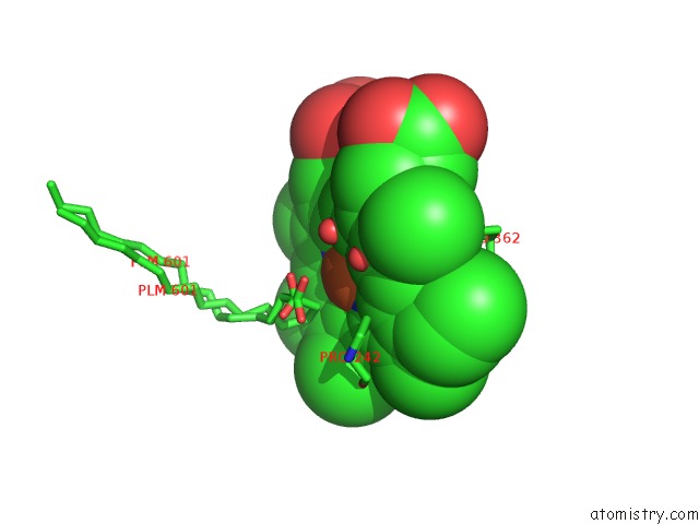
Mono view
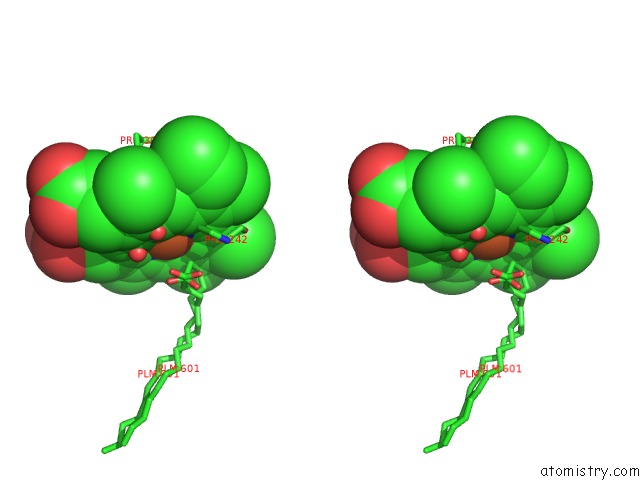
Stereo pair view
Mono view
Stereo pair view
A full contact list of Iron with other atoms in the Fe binding
site number 1 of Cytochrome P450SP Alpha (CYP152B1) Mutant L78F within 5.0Å range:
|
Reference:
T.Fujishiro,
O.Shoji,
S.Nagano,
H.Sugimoto,
Y.Shiro,
Y.Watanabe.
Crystal Structure of H2O2-Dependent Cytochrome P450SPALPHA with Its Bound Fatty Acid Substrate: Insight Into the Regioselective Hydroxylation of Fatty Acids at the Alpha Position. J.Biol.Chem. V. 286 29941 2011.
ISSN: ISSN 0021-9258
PubMed: 21719702
DOI: 10.1074/JBC.M111.245225
Page generated: Mon Aug 4 23:35:08 2025
ISSN: ISSN 0021-9258
PubMed: 21719702
DOI: 10.1074/JBC.M111.245225
Last articles
Mg in 5G15Mg in 5FWP
Mg in 5G0V
Mg in 5FZ5
Mg in 5FWJ
Mg in 5FYW
Mg in 5FWM
Mg in 5FWL
Mg in 5FWK
Mg in 5FV7