Iron »
PDB 3b9j-3by0 »
3ben »
Iron in PDB 3ben: Structure of N-(12-Imidazolyl-Dodecanoyl)-L-Leucine Inhibitor Bound to the Heme Domain of Cytochrome P450-BM3
Enzymatic activity of Structure of N-(12-Imidazolyl-Dodecanoyl)-L-Leucine Inhibitor Bound to the Heme Domain of Cytochrome P450-BM3
All present enzymatic activity of Structure of N-(12-Imidazolyl-Dodecanoyl)-L-Leucine Inhibitor Bound to the Heme Domain of Cytochrome P450-BM3:
1.14.14.1;
1.14.14.1;
Protein crystallography data
The structure of Structure of N-(12-Imidazolyl-Dodecanoyl)-L-Leucine Inhibitor Bound to the Heme Domain of Cytochrome P450-BM3, PDB code: 3ben
was solved by
D.R.Tomchick,
with X-Ray Crystallography technique. A brief refinement statistics is given in the table below:
| Resolution Low / High (Å) | 30.00 / 1.65 |
| Space group | P 1 21 1 |
| Cell size a, b, c (Å), α, β, γ (°) | 58.820, 148.209, 63.792, 90.00, 98.32, 90.00 |
| R / Rfree (%) | 16.1 / 19.1 |
Other elements in 3ben:
The structure of Structure of N-(12-Imidazolyl-Dodecanoyl)-L-Leucine Inhibitor Bound to the Heme Domain of Cytochrome P450-BM3 also contains other interesting chemical elements:
| Magnesium | (Mg) | 1 atom |
Iron Binding Sites:
The binding sites of Iron atom in the Structure of N-(12-Imidazolyl-Dodecanoyl)-L-Leucine Inhibitor Bound to the Heme Domain of Cytochrome P450-BM3
(pdb code 3ben). This binding sites where shown within
5.0 Angstroms radius around Iron atom.
In total 4 binding sites of Iron where determined in the Structure of N-(12-Imidazolyl-Dodecanoyl)-L-Leucine Inhibitor Bound to the Heme Domain of Cytochrome P450-BM3, PDB code: 3ben:
Jump to Iron binding site number: 1; 2; 3; 4;
In total 4 binding sites of Iron where determined in the Structure of N-(12-Imidazolyl-Dodecanoyl)-L-Leucine Inhibitor Bound to the Heme Domain of Cytochrome P450-BM3, PDB code: 3ben:
Jump to Iron binding site number: 1; 2; 3; 4;
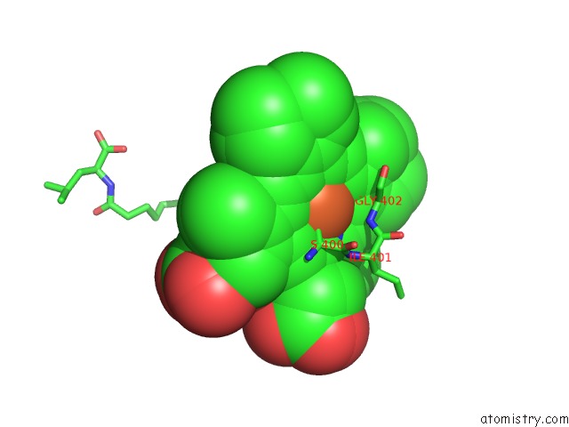
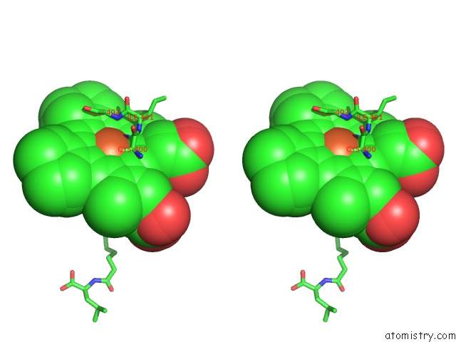
Iron binding site 1 out of 4 in 3ben
Go back to
Iron binding site 1 out
of 4 in the Structure of N-(12-Imidazolyl-Dodecanoyl)-L-Leucine Inhibitor Bound to the Heme Domain of Cytochrome P450-BM3
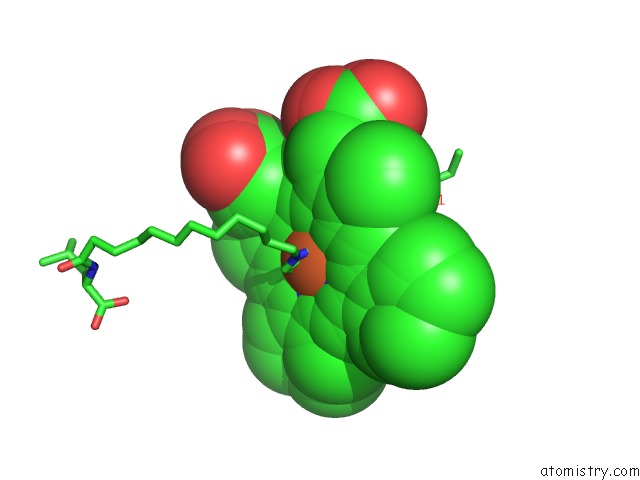
Mono view
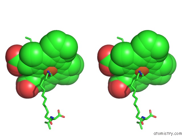
Stereo pair view
Mono view
Stereo pair view
A full contact list of Iron with other atoms in the Fe binding
site number 1 of Structure of N-(12-Imidazolyl-Dodecanoyl)-L-Leucine Inhibitor Bound to the Heme Domain of Cytochrome P450-BM3 within 5.0Å range:
|
Iron binding site 2 out of 4 in 3ben
Go back to
Iron binding site 2 out
of 4 in the Structure of N-(12-Imidazolyl-Dodecanoyl)-L-Leucine Inhibitor Bound to the Heme Domain of Cytochrome P450-BM3
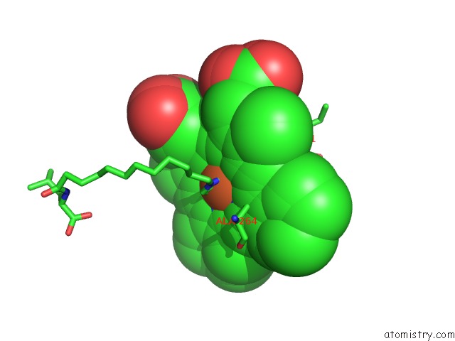
Mono view
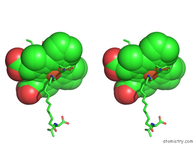
Stereo pair view
Mono view
Stereo pair view
A full contact list of Iron with other atoms in the Fe binding
site number 2 of Structure of N-(12-Imidazolyl-Dodecanoyl)-L-Leucine Inhibitor Bound to the Heme Domain of Cytochrome P450-BM3 within 5.0Å range:
|
Iron binding site 3 out of 4 in 3ben
Go back to
Iron binding site 3 out
of 4 in the Structure of N-(12-Imidazolyl-Dodecanoyl)-L-Leucine Inhibitor Bound to the Heme Domain of Cytochrome P450-BM3
Mono view
Stereo pair view
Mono view
Stereo pair view
A full contact list of Iron with other atoms in the Fe binding
site number 3 of Structure of N-(12-Imidazolyl-Dodecanoyl)-L-Leucine Inhibitor Bound to the Heme Domain of Cytochrome P450-BM3 within 5.0Å range:
|
Iron binding site 4 out of 4 in 3ben
Go back to
Iron binding site 4 out
of 4 in the Structure of N-(12-Imidazolyl-Dodecanoyl)-L-Leucine Inhibitor Bound to the Heme Domain of Cytochrome P450-BM3

Mono view

Stereo pair view
Mono view
Stereo pair view
A full contact list of Iron with other atoms in the Fe binding
site number 4 of Structure of N-(12-Imidazolyl-Dodecanoyl)-L-Leucine Inhibitor Bound to the Heme Domain of Cytochrome P450-BM3 within 5.0Å range:
|
Reference:
D.C.Haines,
B.Chen,
D.R.Tomchick,
M.Bondlela,
A.Hegde,
M.Machius,
J.A.Peterson.
Crystal Structure of Inhibitor-Bound P450BM-3 Reveals Open Conformation of Substrate Access Channel. Biochemistry V. 47 3662 2008.
ISSN: ISSN 0006-2960
PubMed: 18298086
DOI: 10.1021/BI7023964
Page generated: Mon Aug 4 23:52:59 2025
ISSN: ISSN 0006-2960
PubMed: 18298086
DOI: 10.1021/BI7023964
Last articles
Mg in 6ZNDMg in 6ZNW
Mg in 6ZJB
Mg in 6ZN7
Mg in 6ZN4
Mg in 6ZMD
Mg in 6ZLI
Mg in 6ZM2
Mg in 6ZL7
Mg in 6ZL5