Iron »
PDB 3b9j-3by0 »
3bom »
Iron in PDB 3bom: Crystal Structure of Trout Hemoglobin at 1.35 Angstrom Resolution
Protein crystallography data
The structure of Crystal Structure of Trout Hemoglobin at 1.35 Angstrom Resolution, PDB code: 3bom
was solved by
R.Aranda Iv,
C.A.Bingman,
E.Bitto,
G.E.Wesenberg,
M.Richards,
G.N.Phillipsjr.,
Center For Eukaryotic Structural Genomics (Cesg),
with X-Ray Crystallography technique. A brief refinement statistics is given in the table below:
| Resolution Low / High (Å) | 39.29 / 1.35 |
| Space group | P 1 21 1 |
| Cell size a, b, c (Å), α, β, γ (°) | 57.455, 63.162, 78.702, 90.00, 93.10, 90.00 |
| R / Rfree (%) | 17 / 21.2 |
Iron Binding Sites:
The binding sites of Iron atom in the Crystal Structure of Trout Hemoglobin at 1.35 Angstrom Resolution
(pdb code 3bom). This binding sites where shown within
5.0 Angstroms radius around Iron atom.
In total 4 binding sites of Iron where determined in the Crystal Structure of Trout Hemoglobin at 1.35 Angstrom Resolution, PDB code: 3bom:
Jump to Iron binding site number: 1; 2; 3; 4;
In total 4 binding sites of Iron where determined in the Crystal Structure of Trout Hemoglobin at 1.35 Angstrom Resolution, PDB code: 3bom:
Jump to Iron binding site number: 1; 2; 3; 4;
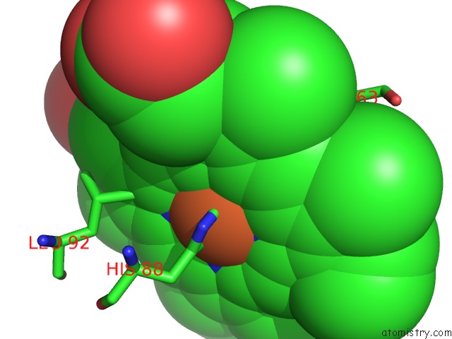
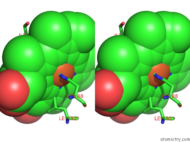
Iron binding site 1 out of 4 in 3bom
Go back to
Iron binding site 1 out
of 4 in the Crystal Structure of Trout Hemoglobin at 1.35 Angstrom Resolution
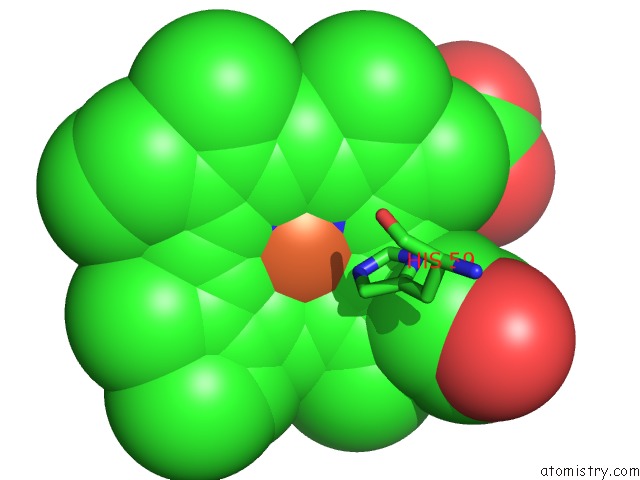
Mono view
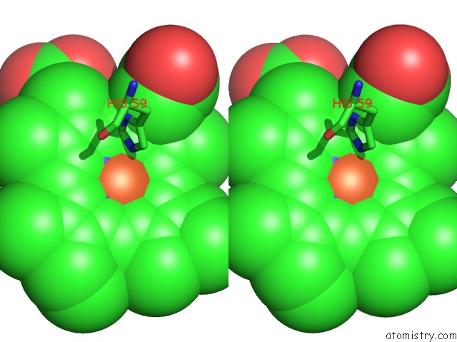
Stereo pair view
Mono view
Stereo pair view
A full contact list of Iron with other atoms in the Fe binding
site number 1 of Crystal Structure of Trout Hemoglobin at 1.35 Angstrom Resolution within 5.0Å range:
|
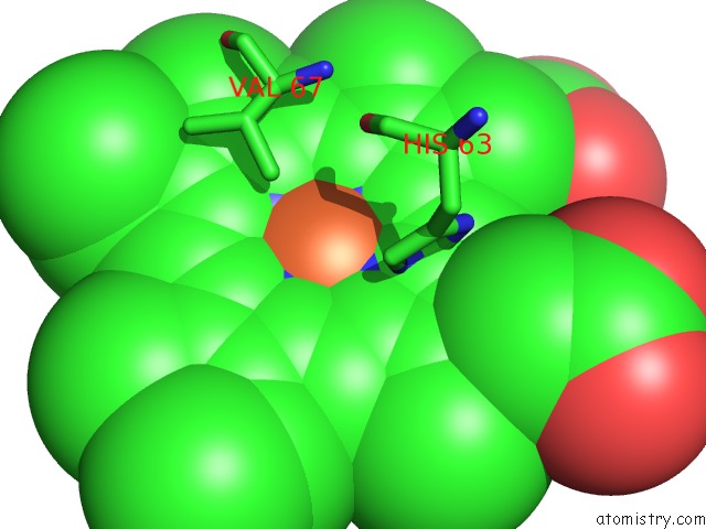
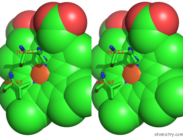
Iron binding site 2 out of 4 in 3bom
Go back to
Iron binding site 2 out
of 4 in the Crystal Structure of Trout Hemoglobin at 1.35 Angstrom Resolution
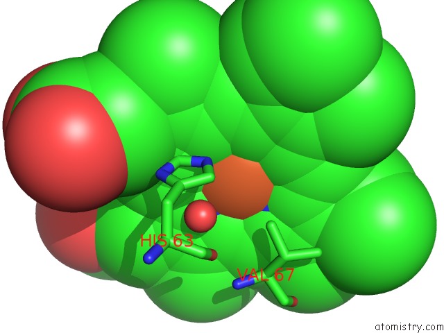
Mono view
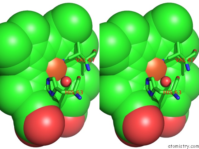
Stereo pair view
Mono view
Stereo pair view
A full contact list of Iron with other atoms in the Fe binding
site number 2 of Crystal Structure of Trout Hemoglobin at 1.35 Angstrom Resolution within 5.0Å range:
|
Iron binding site 3 out of 4 in 3bom
Go back to
Iron binding site 3 out
of 4 in the Crystal Structure of Trout Hemoglobin at 1.35 Angstrom Resolution
Mono view
Stereo pair view
Mono view
Stereo pair view
A full contact list of Iron with other atoms in the Fe binding
site number 3 of Crystal Structure of Trout Hemoglobin at 1.35 Angstrom Resolution within 5.0Å range:
|
Iron binding site 4 out of 4 in 3bom
Go back to
Iron binding site 4 out
of 4 in the Crystal Structure of Trout Hemoglobin at 1.35 Angstrom Resolution
Mono view
Stereo pair view
Mono view
Stereo pair view
A full contact list of Iron with other atoms in the Fe binding
site number 4 of Crystal Structure of Trout Hemoglobin at 1.35 Angstrom Resolution within 5.0Å range:
|
Reference:
R.Aranda Iv,
C.A.Bingman,
E.Bitto,
G.E.Wesenberg,
M.Richards,
G.N.Phillips Jr..
Trout Hemoglobin Crystal Structure at 1.35 Angstroms Resolution. To Be Published.
Page generated: Mon Aug 4 23:59:45 2025
Last articles
Xe in 2OQUXe in 2IE6
Xe in 2IC0
Xe in 2DKI
Xe in 2FIC
Xe in 2A7A
Xe in 2A7D
Xe in 2A9R
Xe in 2A7B
Xe in 1ZDM