Iron »
PDB 3c25-3crb »
3c78 »
Iron in PDB 3c78: 0.98 A Crystal Structure of Nitrophorin 4 From Rhodnius Prolixus Containing Fe(III) 2,4 Dimethyl Deuteroporphyrin IX Complexed with Ammonia at pH 7.5
Protein crystallography data
The structure of 0.98 A Crystal Structure of Nitrophorin 4 From Rhodnius Prolixus Containing Fe(III) 2,4 Dimethyl Deuteroporphyrin IX Complexed with Ammonia at pH 7.5, PDB code: 3c78
was solved by
A.M.Amoia,
W.R.Montfort,
with X-Ray Crystallography technique. A brief refinement statistics is given in the table below:
| Resolution Low / High (Å) | 20.30 / 0.98 |
| Space group | C 1 2 1 |
| Cell size a, b, c (Å), α, β, γ (°) | 69.873, 42.490, 52.633, 90.00, 94.08, 90.00 |
| R / Rfree (%) | 16.5 / 18.9 |
Iron Binding Sites:
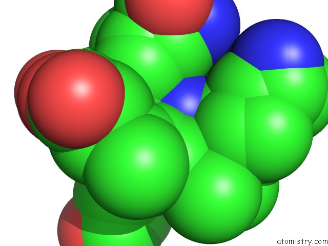
The binding sites of Iron atom in the 0.98 A Crystal Structure of Nitrophorin 4 From Rhodnius Prolixus Containing Fe(III) 2,4 Dimethyl Deuteroporphyrin IX Complexed with Ammonia at pH 7.5
(pdb code 3c78). This binding sites where shown within
5.0 Angstroms radius around Iron atom.
In total only one binding site of Iron was determined in the 0.98 A Crystal Structure of Nitrophorin 4 From Rhodnius Prolixus Containing Fe(III) 2,4 Dimethyl Deuteroporphyrin IX Complexed with Ammonia at pH 7.5, PDB code: 3c78:
In total only one binding site of Iron was determined in the 0.98 A Crystal Structure of Nitrophorin 4 From Rhodnius Prolixus Containing Fe(III) 2,4 Dimethyl Deuteroporphyrin IX Complexed with Ammonia at pH 7.5, PDB code: 3c78:
Iron binding site 1 out of 1 in 3c78
Go back to
Iron binding site 1 out
of 1 in the 0.98 A Crystal Structure of Nitrophorin 4 From Rhodnius Prolixus Containing Fe(III) 2,4 Dimethyl Deuteroporphyrin IX Complexed with Ammonia at pH 7.5
Mono view

Stereo pair view
Mono view
Stereo pair view
A full contact list of Iron with other atoms in the Fe binding
site number 1 of 0.98 A Crystal Structure of Nitrophorin 4 From Rhodnius Prolixus Containing Fe(III) 2,4 Dimethyl Deuteroporphyrin IX Complexed with Ammonia at pH 7.5 within 5.0Å range:
|
Reference:
A.M.Amoia,
W.R.Montfort.
Heme Distortion in Nitrophorin 4: High Resolution Structures of Mutated Positions L123V and L133V and Heme Altered Proteins To Be Published.
Page generated: Sun Aug 4 08:15:11 2024
Last articles
Fe in 2YXOFe in 2YRS
Fe in 2YXC
Fe in 2YNM
Fe in 2YVJ
Fe in 2YP1
Fe in 2YU2
Fe in 2YU1
Fe in 2YQB
Fe in 2YOO