Iron »
PDB 3crv-3dby »
3d89 »
Iron in PDB 3d89: Crystal Structure of A Soluble Rieske Ferredoxin From Mus Musculus
Protein crystallography data
The structure of Crystal Structure of A Soluble Rieske Ferredoxin From Mus Musculus, PDB code: 3d89
was solved by
E.J.Levin,
J.G.Mccoy,
N.L.Elsen,
K.D.Seder,
C.A.Bingman,
G.E.Wesenberg,
B.G.Fox,
G.N.Phillips Jr.,
Center For Eukaryotic Structural Genomics(Cesg),
with X-Ray Crystallography technique. A brief refinement statistics is given in the table below:
| Resolution Low / High (Å) | 37.75 / 2.07 |
| Space group | P 43 21 2 |
| Cell size a, b, c (Å), α, β, γ (°) | 52.413, 52.413, 108.808, 90.00, 90.00, 90.00 |
| R / Rfree (%) | 19.9 / 22.8 |
Iron Binding Sites:
The binding sites of Iron atom in the Crystal Structure of A Soluble Rieske Ferredoxin From Mus Musculus
(pdb code 3d89). This binding sites where shown within
5.0 Angstroms radius around Iron atom.
In total 2 binding sites of Iron where determined in the Crystal Structure of A Soluble Rieske Ferredoxin From Mus Musculus, PDB code: 3d89:
Jump to Iron binding site number: 1; 2;
In total 2 binding sites of Iron where determined in the Crystal Structure of A Soluble Rieske Ferredoxin From Mus Musculus, PDB code: 3d89:
Jump to Iron binding site number: 1; 2;
Iron binding site 1 out of 2 in 3d89
Go back to
Iron binding site 1 out
of 2 in the Crystal Structure of A Soluble Rieske Ferredoxin From Mus Musculus
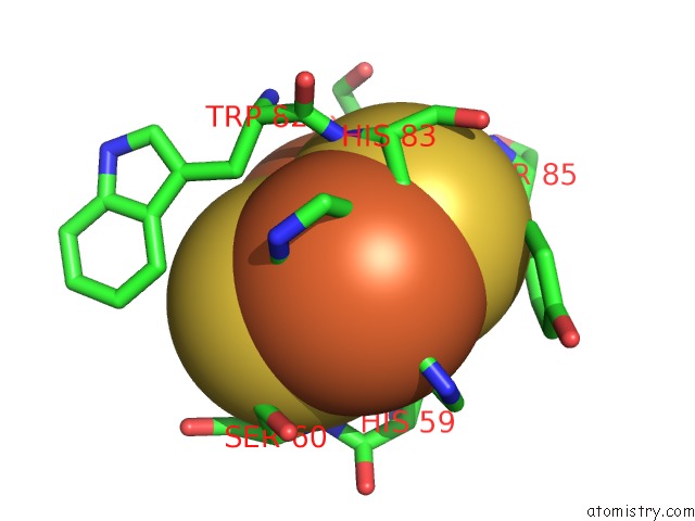
Mono view
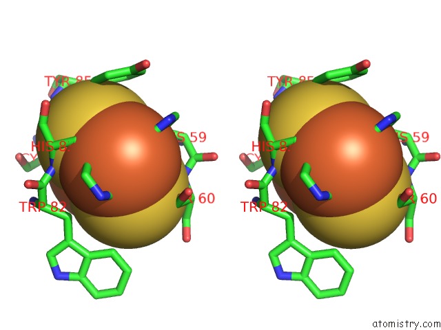
Stereo pair view
Mono view
Stereo pair view
A full contact list of Iron with other atoms in the Fe binding
site number 1 of Crystal Structure of A Soluble Rieske Ferredoxin From Mus Musculus within 5.0Å range:
|
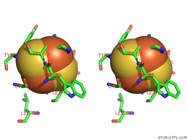
Iron binding site 2 out of 2 in 3d89
Go back to
Iron binding site 2 out
of 2 in the Crystal Structure of A Soluble Rieske Ferredoxin From Mus Musculus
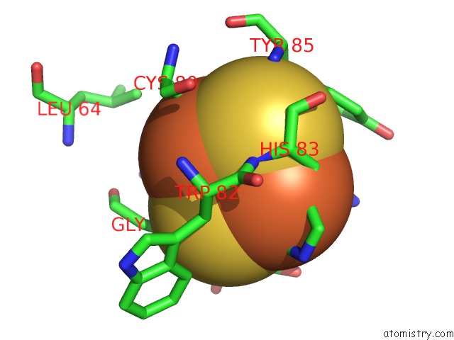
Mono view
Stereo pair view
Mono view
Stereo pair view
A full contact list of Iron with other atoms in the Fe binding
site number 2 of Crystal Structure of A Soluble Rieske Ferredoxin From Mus Musculus within 5.0Å range:
|
Reference:
E.J.Levin,
N.L.Elsen,
K.D.Seder,
J.G.Mccoy,
B.G.Fox,
G.N.Phillips.
X-Ray Structure of A Soluble Rieske-Type Ferredoxin From Mus Musculus. Acta Crystallogr.,Sect.D V. 64 933 2008.
ISSN: ISSN 0907-4449
PubMed: 18703841
DOI: 10.1107/S0907444908021653
Page generated: Tue Aug 5 00:31:49 2025
ISSN: ISSN 0907-4449
PubMed: 18703841
DOI: 10.1107/S0907444908021653
Last articles
W in 9FPPW in 8PRM
W in 9QM1
W in 9QM0
W in 9OJ3
W in 9MQX
W in 9FP4
W in 9BEO
W in 9BEM
W in 8P2U