Iron »
PDB 3dcp-3e0f »
3dhh »
Iron in PDB 3dhh: Crystal Structure of Resting State Toluene 4-Monoxygenase Hydroxylase Complexed with Effector Protein
Protein crystallography data
The structure of Crystal Structure of Resting State Toluene 4-Monoxygenase Hydroxylase Complexed with Effector Protein, PDB code: 3dhh
was solved by
L.J.Bailey,
J.G.Mccoy,
G.N.Phillips Jr.,
B.G.Fox,
with X-Ray Crystallography technique. A brief refinement statistics is given in the table below:
| Resolution Low / High (Å) | 91.29 / 1.94 |
| Space group | C 2 2 21 |
| Cell size a, b, c (Å), α, β, γ (°) | 100.418, 115.613, 182.403, 90.00, 90.00, 90.00 |
| R / Rfree (%) | 15.7 / 20.2 |
Other elements in 3dhh:
The structure of Crystal Structure of Resting State Toluene 4-Monoxygenase Hydroxylase Complexed with Effector Protein also contains other interesting chemical elements:
| Bromine | (Br) | 5 atoms |
| Chlorine | (Cl) | 1 atom |
Iron Binding Sites:
The binding sites of Iron atom in the Crystal Structure of Resting State Toluene 4-Monoxygenase Hydroxylase Complexed with Effector Protein
(pdb code 3dhh). This binding sites where shown within
5.0 Angstroms radius around Iron atom.
In total 2 binding sites of Iron where determined in the Crystal Structure of Resting State Toluene 4-Monoxygenase Hydroxylase Complexed with Effector Protein, PDB code: 3dhh:
Jump to Iron binding site number: 1; 2;
In total 2 binding sites of Iron where determined in the Crystal Structure of Resting State Toluene 4-Monoxygenase Hydroxylase Complexed with Effector Protein, PDB code: 3dhh:
Jump to Iron binding site number: 1; 2;
Iron binding site 1 out of 2 in 3dhh
Go back to
Iron binding site 1 out
of 2 in the Crystal Structure of Resting State Toluene 4-Monoxygenase Hydroxylase Complexed with Effector Protein
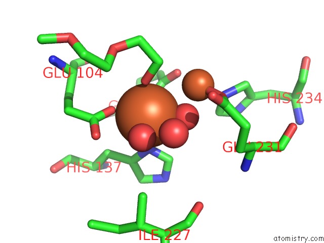
Mono view
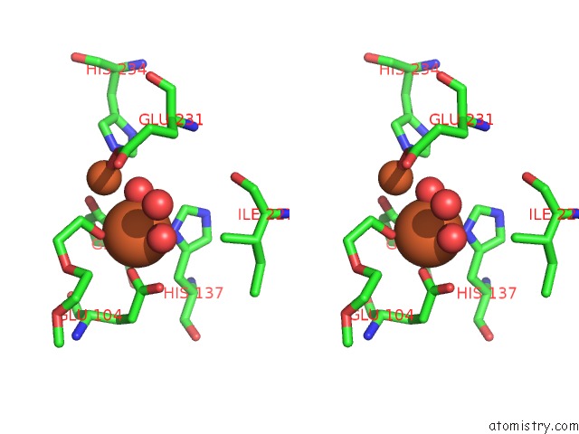
Stereo pair view
Mono view
Stereo pair view
A full contact list of Iron with other atoms in the Fe binding
site number 1 of Crystal Structure of Resting State Toluene 4-Monoxygenase Hydroxylase Complexed with Effector Protein within 5.0Å range:
|
Iron binding site 2 out of 2 in 3dhh
Go back to
Iron binding site 2 out
of 2 in the Crystal Structure of Resting State Toluene 4-Monoxygenase Hydroxylase Complexed with Effector Protein
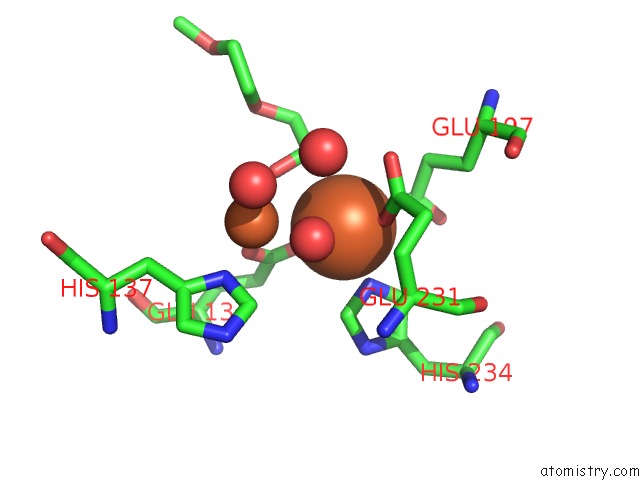
Mono view
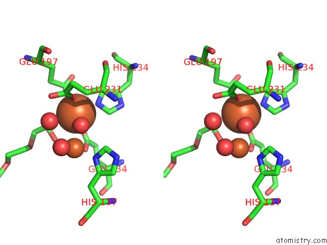
Stereo pair view
Mono view
Stereo pair view
A full contact list of Iron with other atoms in the Fe binding
site number 2 of Crystal Structure of Resting State Toluene 4-Monoxygenase Hydroxylase Complexed with Effector Protein within 5.0Å range:
|
Reference:
L.J.Bailey,
J.G.Mccoy,
G.N.Phillips Jr.,
B.G.Fox.
Structural Consequences of Effector Protein Complex Formation in A Diiron Hydroxylase. Proc.Natl.Acad.Sci.Usa V. 105 19194 2008.
ISSN: ISSN 0027-8424
PubMed: 19033467
DOI: 10.1073/PNAS.0807948105
Page generated: Tue Aug 5 00:37:10 2025
ISSN: ISSN 0027-8424
PubMed: 19033467
DOI: 10.1073/PNAS.0807948105
Last articles
K in 8EVXK in 8EH1
K in 8EH0
K in 8EGZ
K in 8EGY
K in 8EFF
K in 8EGF
K in 8EGD
K in 8EDP
K in 8EBO