Iron »
PDB 3dcp-3e0f »
3du2 »
Iron in PDB 3du2: E(L212)A Mutant Structure of Photosynthetic Reaction Center From Rhodobacter Sphaeroides
Protein crystallography data
The structure of E(L212)A Mutant Structure of Photosynthetic Reaction Center From Rhodobacter Sphaeroides, PDB code: 3du2
was solved by
P.R.Pokkuluri,
M.Schiffer,
with X-Ray Crystallography technique. A brief refinement statistics is given in the table below:
| Resolution Low / High (Å) | 30.00 / 3.10 |
| Space group | P 31 2 1 |
| Cell size a, b, c (Å), α, β, γ (°) | 141.600, 141.600, 187.300, 90.00, 90.00, 120.00 |
| R / Rfree (%) | 19.2 / 19.5 |
Other elements in 3du2:
The structure of E(L212)A Mutant Structure of Photosynthetic Reaction Center From Rhodobacter Sphaeroides also contains other interesting chemical elements:
| Magnesium | (Mg) | 4 atoms |
Iron Binding Sites:
The binding sites of Iron atom in the E(L212)A Mutant Structure of Photosynthetic Reaction Center From Rhodobacter Sphaeroides
(pdb code 3du2). This binding sites where shown within
5.0 Angstroms radius around Iron atom.
In total only one binding site of Iron was determined in the E(L212)A Mutant Structure of Photosynthetic Reaction Center From Rhodobacter Sphaeroides, PDB code: 3du2:
In total only one binding site of Iron was determined in the E(L212)A Mutant Structure of Photosynthetic Reaction Center From Rhodobacter Sphaeroides, PDB code: 3du2:
Iron binding site 1 out of 1 in 3du2
Go back to
Iron binding site 1 out
of 1 in the E(L212)A Mutant Structure of Photosynthetic Reaction Center From Rhodobacter Sphaeroides
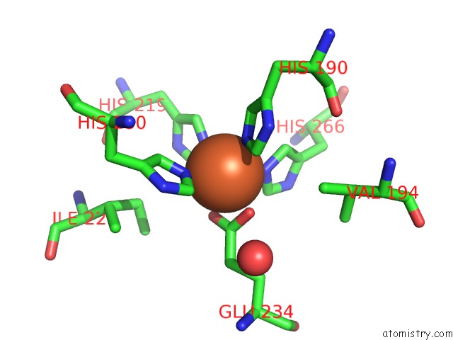
Mono view
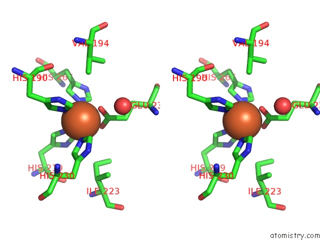
Stereo pair view
Mono view
Stereo pair view
A full contact list of Iron with other atoms in the Fe binding
site number 1 of E(L212)A Mutant Structure of Photosynthetic Reaction Center From Rhodobacter Sphaeroides within 5.0Å range:
|
Reference:
P.R.Pokkuluri,
P.D.Laible,
D.K.Hanson,
M.Schiffer.
E(L212)A Mutant Structure of Photosynthetic Reaction Center From Rhodobacter Sphaeroides To Be Published.
Page generated: Tue Aug 5 00:41:16 2025
Last articles
Fe in 5TH5Fe in 5T5M
Fe in 5TIA
Fe in 5TI9
Fe in 5TGS
Fe in 5TFU
Fe in 5TG0
Fe in 5TFT
Fe in 5TE8
Fe in 5TDV