Iron »
PDB 3e13-3eai »
3e13 »
Iron in PDB 3e13: Iron Reconstituted Ferric Binding Protein From Campylobacter Jejuni
Protein crystallography data
The structure of Iron Reconstituted Ferric Binding Protein From Campylobacter Jejuni, PDB code: 3e13
was solved by
M.E.P.Murphy,
S.A.L.Tom-Yew,
E.G.Bekker,
with X-Ray Crystallography technique. A brief refinement statistics is given in the table below:
| Resolution Low / High (Å) | 48.80 / 1.60 |
| Space group | P 21 21 21 |
| Cell size a, b, c (Å), α, β, γ (°) | 57.113, 54.740, 92.237, 90.00, 90.00, 90.00 |
| R / Rfree (%) | 14.5 / 18.6 |
Iron Binding Sites:
The binding sites of Iron atom in the Iron Reconstituted Ferric Binding Protein From Campylobacter Jejuni
(pdb code 3e13). This binding sites where shown within
5.0 Angstroms radius around Iron atom.
In total only one binding site of Iron was determined in the Iron Reconstituted Ferric Binding Protein From Campylobacter Jejuni, PDB code: 3e13:
In total only one binding site of Iron was determined in the Iron Reconstituted Ferric Binding Protein From Campylobacter Jejuni, PDB code: 3e13:
Iron binding site 1 out of 1 in 3e13
Go back to
Iron binding site 1 out
of 1 in the Iron Reconstituted Ferric Binding Protein From Campylobacter Jejuni
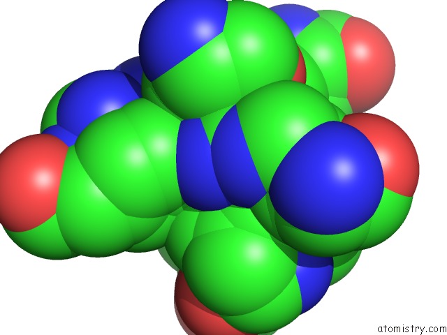
Mono view
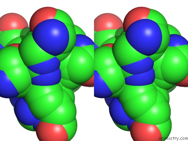
Stereo pair view
Mono view
Stereo pair view
A full contact list of Iron with other atoms in the Fe binding
site number 1 of Iron Reconstituted Ferric Binding Protein From Campylobacter Jejuni within 5.0Å range:
|
Reference:
S.A.L.Tom-Yew,
B.H.Shilton,
E.G.Bekker,
E.C.Gaynor,
M.E.P.Murphy.
Small Hinge Motion By the Anion-Independent Ferric Binding Protein From Campylobacter Jejuni To Be Published.
Page generated: Tue Aug 5 00:43:22 2025
Last articles
Zn in 1SATZn in 1SA5
Zn in 1SA4
Zn in 1S9Z
Zn in 1S7D
Zn in 1S64
Zn in 1S03
Zn in 1S4I
Zn in 1S63
Zn in 1S5P