Iron »
PDB 3e13-3eai »
3e1q »
Iron in PDB 3e1q: Crystal Structure of W133F Variant E. Coli Bacterioferritn with Iron.
Protein crystallography data
The structure of Crystal Structure of W133F Variant E. Coli Bacterioferritn with Iron., PDB code: 3e1q
was solved by
A.Crow,
T.L.Lawson,
A.Lewin,
G.R.Moore,
N.E.Le Brun,
with X-Ray Crystallography technique. A brief refinement statistics is given in the table below:
| Resolution Low / High (Å) | 33.31 / 2.60 |
| Space group | P 42 21 2 |
| Cell size a, b, c (Å), α, β, γ (°) | 207.659, 207.659, 142.767, 90.00, 90.00, 90.00 |
| R / Rfree (%) | 24.5 / 26.3 |
Iron Binding Sites:
Pages:
>>> Page 1 <<< Page 2, Binding sites: 11 - 20; Page 3, Binding sites: 21 - 30;Binding sites:
The binding sites of Iron atom in the Crystal Structure of W133F Variant E. Coli Bacterioferritn with Iron. (pdb code 3e1q). This binding sites where shown within 5.0 Angstroms radius around Iron atom.In total 30 binding sites of Iron where determined in the Crystal Structure of W133F Variant E. Coli Bacterioferritn with Iron., PDB code: 3e1q:
Jump to Iron binding site number: 1; 2; 3; 4; 5; 6; 7; 8; 9; 10;
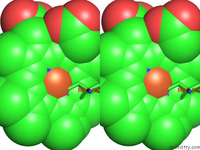
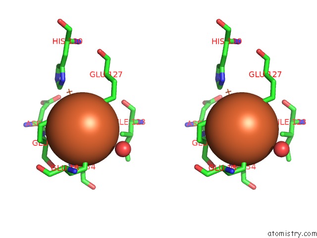
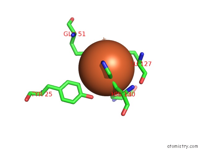
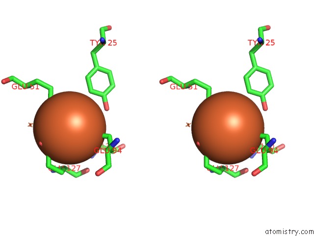
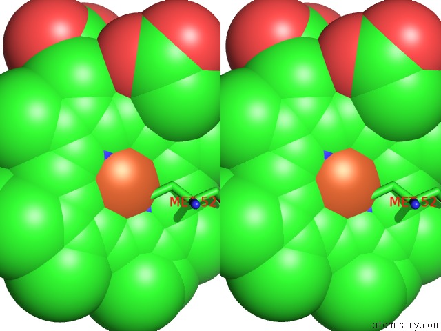
Iron binding site 1 out of 30 in 3e1q
Go back to
Iron binding site 1 out
of 30 in the Crystal Structure of W133F Variant E. Coli Bacterioferritn with Iron.
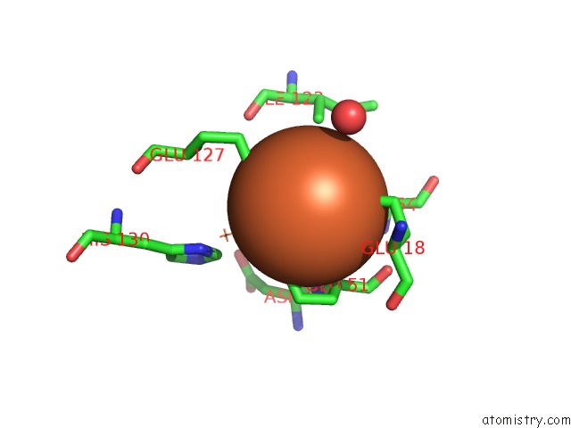
Mono view

Stereo pair view
Mono view
Stereo pair view
A full contact list of Iron with other atoms in the Fe binding
site number 1 of Crystal Structure of W133F Variant E. Coli Bacterioferritn with Iron. within 5.0Å range:
|
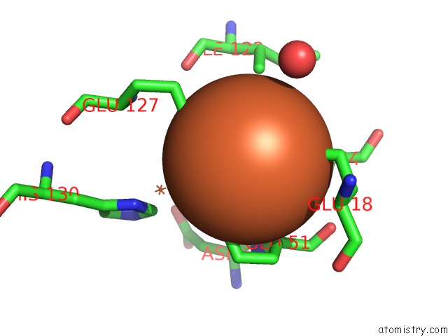
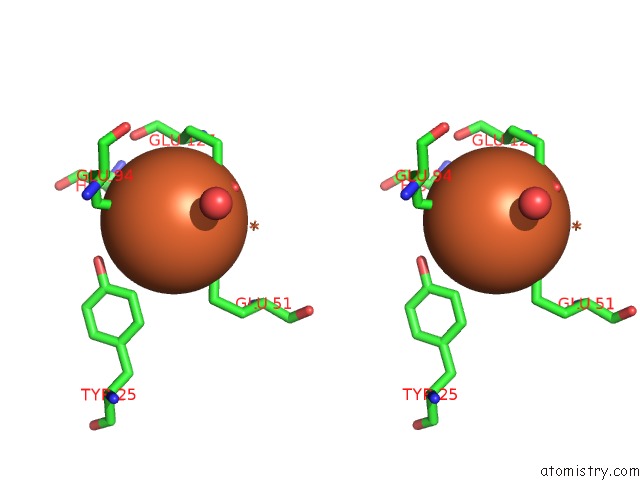
Iron binding site 2 out of 30 in 3e1q
Go back to
Iron binding site 2 out
of 30 in the Crystal Structure of W133F Variant E. Coli Bacterioferritn with Iron.
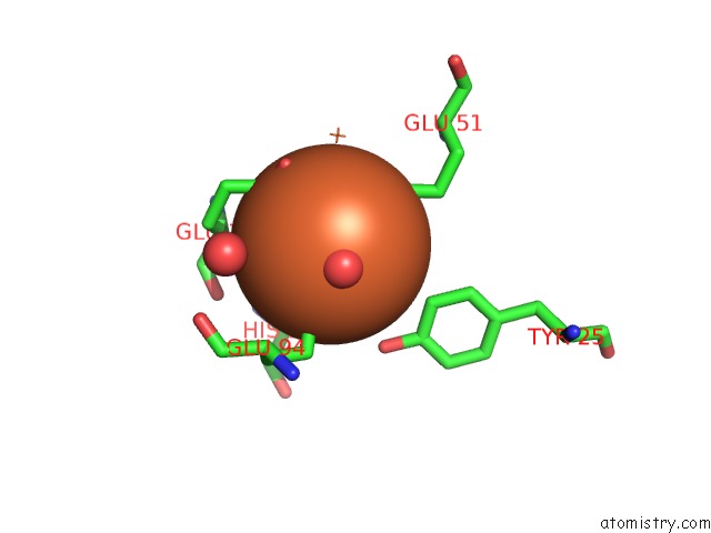
Mono view

Stereo pair view
Mono view
Stereo pair view
A full contact list of Iron with other atoms in the Fe binding
site number 2 of Crystal Structure of W133F Variant E. Coli Bacterioferritn with Iron. within 5.0Å range:
|
Iron binding site 3 out of 30 in 3e1q
Go back to
Iron binding site 3 out
of 30 in the Crystal Structure of W133F Variant E. Coli Bacterioferritn with Iron.

Mono view
Stereo pair view
Mono view
Stereo pair view
A full contact list of Iron with other atoms in the Fe binding
site number 3 of Crystal Structure of W133F Variant E. Coli Bacterioferritn with Iron. within 5.0Å range:
|
Iron binding site 4 out of 30 in 3e1q
Go back to
Iron binding site 4 out
of 30 in the Crystal Structure of W133F Variant E. Coli Bacterioferritn with Iron.

Mono view
Stereo pair view
Mono view
Stereo pair view
A full contact list of Iron with other atoms in the Fe binding
site number 4 of Crystal Structure of W133F Variant E. Coli Bacterioferritn with Iron. within 5.0Å range:
|
Iron binding site 5 out of 30 in 3e1q
Go back to
Iron binding site 5 out
of 30 in the Crystal Structure of W133F Variant E. Coli Bacterioferritn with Iron.
Mono view

Stereo pair view
Mono view
Stereo pair view
A full contact list of Iron with other atoms in the Fe binding
site number 5 of Crystal Structure of W133F Variant E. Coli Bacterioferritn with Iron. within 5.0Å range:
|
Iron binding site 6 out of 30 in 3e1q
Go back to
Iron binding site 6 out
of 30 in the Crystal Structure of W133F Variant E. Coli Bacterioferritn with Iron.
Mono view

Stereo pair view
Mono view
Stereo pair view
A full contact list of Iron with other atoms in the Fe binding
site number 6 of Crystal Structure of W133F Variant E. Coli Bacterioferritn with Iron. within 5.0Å range:
|
Iron binding site 7 out of 30 in 3e1q
Go back to
Iron binding site 7 out
of 30 in the Crystal Structure of W133F Variant E. Coli Bacterioferritn with Iron.

Mono view
Stereo pair view
Mono view
Stereo pair view
A full contact list of Iron with other atoms in the Fe binding
site number 7 of Crystal Structure of W133F Variant E. Coli Bacterioferritn with Iron. within 5.0Å range:
|
Iron binding site 8 out of 30 in 3e1q
Go back to
Iron binding site 8 out
of 30 in the Crystal Structure of W133F Variant E. Coli Bacterioferritn with Iron.

Mono view

Stereo pair view
Mono view
Stereo pair view
A full contact list of Iron with other atoms in the Fe binding
site number 8 of Crystal Structure of W133F Variant E. Coli Bacterioferritn with Iron. within 5.0Å range:
|
Iron binding site 9 out of 30 in 3e1q
Go back to
Iron binding site 9 out
of 30 in the Crystal Structure of W133F Variant E. Coli Bacterioferritn with Iron.

Mono view
Stereo pair view
Mono view
Stereo pair view
A full contact list of Iron with other atoms in the Fe binding
site number 9 of Crystal Structure of W133F Variant E. Coli Bacterioferritn with Iron. within 5.0Å range:
|
Iron binding site 10 out of 30 in 3e1q
Go back to
Iron binding site 10 out
of 30 in the Crystal Structure of W133F Variant E. Coli Bacterioferritn with Iron.

Mono view
Stereo pair view
Mono view
Stereo pair view
A full contact list of Iron with other atoms in the Fe binding
site number 10 of Crystal Structure of W133F Variant E. Coli Bacterioferritn with Iron. within 5.0Å range:
|
Reference:
T.L.Lawson,
A.Crow,
A.Lewin,
S.Yasmin,
G.R.Moore,
N.E.Le Brun.
Monitoring the Iron Status of the Ferroxidase Center of Escherichia Coli Bacterioferritin Using Fluorescence Spectroscopy. Biochemistry V. 48 9031 2009.
ISSN: ISSN 0006-2960
PubMed: 19705876
DOI: 10.1021/BI900869X
Page generated: Tue Aug 5 00:46:59 2025
ISSN: ISSN 0006-2960
PubMed: 19705876
DOI: 10.1021/BI900869X
Last articles
Zn in 1K81Zn in 1K7Q
Zn in 1K7G
Zn in 1K7I
Zn in 1K53
Zn in 1K52
Zn in 1K1D
Zn in 1K4P
Zn in 1K51
Zn in 1K4H