Iron »
PDB 3e13-3eai »
3e5j »
Iron in PDB 3e5j: Crystal Structure of CYP105P1 Wild-Type Ligand-Free Form
Protein crystallography data
The structure of Crystal Structure of CYP105P1 Wild-Type Ligand-Free Form, PDB code: 3e5j
was solved by
L.H.Xu,
S.Fushinobu,
H.Ikeda,
T.Wakagi,
H.Shoun,
with X-Ray Crystallography technique. A brief refinement statistics is given in the table below:
| Resolution Low / High (Å) | 38.75 / 1.95 |
| Space group | P 43 21 2 |
| Cell size a, b, c (Å), α, β, γ (°) | 88.401, 88.401, 193.885, 90.00, 90.00, 90.00 |
| R / Rfree (%) | 19 / 21.1 |
Iron Binding Sites:
The binding sites of Iron atom in the Crystal Structure of CYP105P1 Wild-Type Ligand-Free Form
(pdb code 3e5j). This binding sites where shown within
5.0 Angstroms radius around Iron atom.
In total only one binding site of Iron was determined in the Crystal Structure of CYP105P1 Wild-Type Ligand-Free Form, PDB code: 3e5j:
In total only one binding site of Iron was determined in the Crystal Structure of CYP105P1 Wild-Type Ligand-Free Form, PDB code: 3e5j:
Iron binding site 1 out of 1 in 3e5j
Go back to
Iron binding site 1 out
of 1 in the Crystal Structure of CYP105P1 Wild-Type Ligand-Free Form
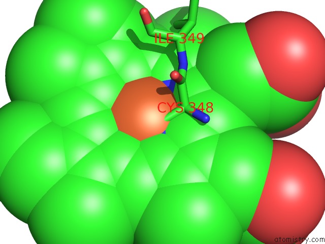
Mono view
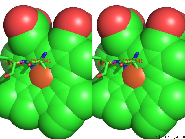
Stereo pair view
Mono view
Stereo pair view
A full contact list of Iron with other atoms in the Fe binding
site number 1 of Crystal Structure of CYP105P1 Wild-Type Ligand-Free Form within 5.0Å range:
|
Reference:
L.H.Xu,
S.Fushinobu,
H.Ikeda,
T.Wakagi,
H.Shoun.
Crystal Structures of Cytochrome P450 105P1 From Streptomyces Avermitilis: Conformational Flexibility and Histidine Ligation State J.Bacteriol. V. 191 1211 2009.
ISSN: ISSN 0021-9193
PubMed: 19074393
DOI: 10.1128/JB.01276-08
Page generated: Tue Aug 5 00:51:48 2025
ISSN: ISSN 0021-9193
PubMed: 19074393
DOI: 10.1128/JB.01276-08
Last articles
Zn in 1K7GZn in 1K7I
Zn in 1K53
Zn in 1K52
Zn in 1K1D
Zn in 1K4P
Zn in 1K51
Zn in 1K4H
Zn in 1K4G
Zn in 1K3X