Iron »
PDB 3ebd-3esf »
3ecj »
Iron in PDB 3ecj: Structure of E323L Mutant of Homoprotocatechuate 2,3-Dioxygenase From Brevibacterium Fuscum at 1.65A Resolution
Enzymatic activity of Structure of E323L Mutant of Homoprotocatechuate 2,3-Dioxygenase From Brevibacterium Fuscum at 1.65A Resolution
All present enzymatic activity of Structure of E323L Mutant of Homoprotocatechuate 2,3-Dioxygenase From Brevibacterium Fuscum at 1.65A Resolution:
1.13.11.15;
1.13.11.15;
Protein crystallography data
The structure of Structure of E323L Mutant of Homoprotocatechuate 2,3-Dioxygenase From Brevibacterium Fuscum at 1.65A Resolution, PDB code: 3ecj
was solved by
E.G.Kovaleva,
J.D.Lipscomb,
with X-Ray Crystallography technique. A brief refinement statistics is given in the table below:
| Resolution Low / High (Å) | 23.46 / 1.65 |
| Space group | P 21 21 2 |
| Cell size a, b, c (Å), α, β, γ (°) | 110.711, 163.403, 101.584, 90.00, 90.00, 90.00 |
| R / Rfree (%) | 16.9 / 19.4 |
Other elements in 3ecj:
The structure of Structure of E323L Mutant of Homoprotocatechuate 2,3-Dioxygenase From Brevibacterium Fuscum at 1.65A Resolution also contains other interesting chemical elements:
| Chlorine | (Cl) | 4 atoms |
| Calcium | (Ca) | 1 atom |
Iron Binding Sites:
The binding sites of Iron atom in the Structure of E323L Mutant of Homoprotocatechuate 2,3-Dioxygenase From Brevibacterium Fuscum at 1.65A Resolution
(pdb code 3ecj). This binding sites where shown within
5.0 Angstroms radius around Iron atom.
In total 4 binding sites of Iron where determined in the Structure of E323L Mutant of Homoprotocatechuate 2,3-Dioxygenase From Brevibacterium Fuscum at 1.65A Resolution, PDB code: 3ecj:
Jump to Iron binding site number: 1; 2; 3; 4;
In total 4 binding sites of Iron where determined in the Structure of E323L Mutant of Homoprotocatechuate 2,3-Dioxygenase From Brevibacterium Fuscum at 1.65A Resolution, PDB code: 3ecj:
Jump to Iron binding site number: 1; 2; 3; 4;
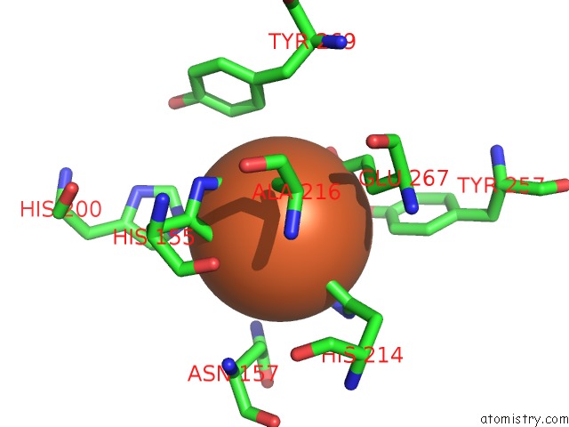
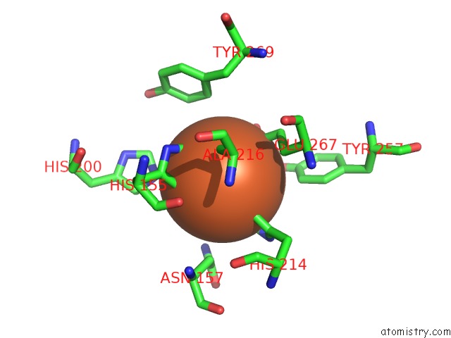
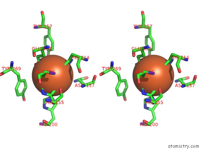
Iron binding site 1 out of 4 in 3ecj
Go back to
Iron binding site 1 out
of 4 in the Structure of E323L Mutant of Homoprotocatechuate 2,3-Dioxygenase From Brevibacterium Fuscum at 1.65A Resolution
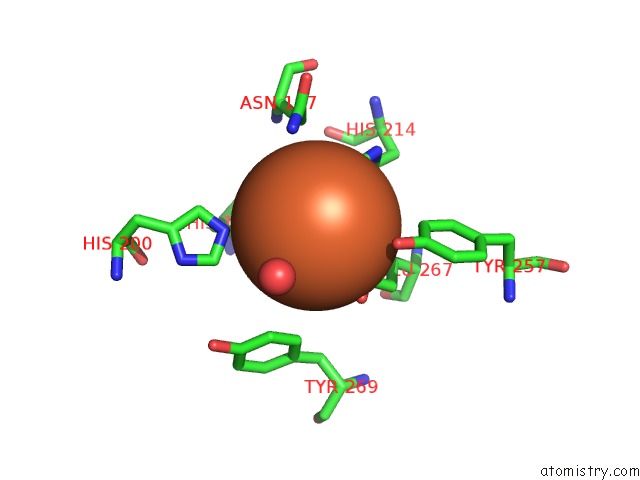
Mono view
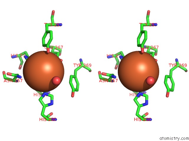
Stereo pair view
Mono view
Stereo pair view
A full contact list of Iron with other atoms in the Fe binding
site number 1 of Structure of E323L Mutant of Homoprotocatechuate 2,3-Dioxygenase From Brevibacterium Fuscum at 1.65A Resolution within 5.0Å range:
|
Iron binding site 2 out of 4 in 3ecj
Go back to
Iron binding site 2 out
of 4 in the Structure of E323L Mutant of Homoprotocatechuate 2,3-Dioxygenase From Brevibacterium Fuscum at 1.65A Resolution
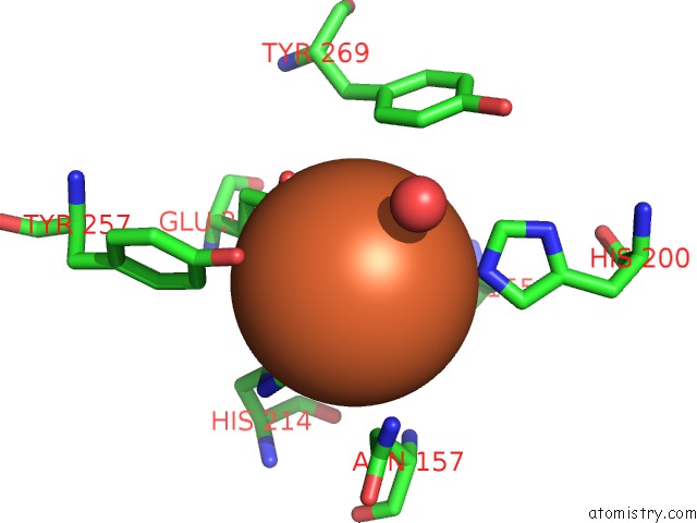
Mono view
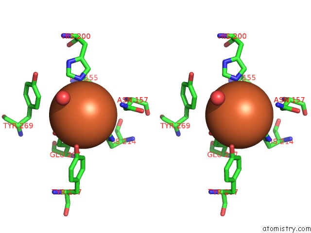
Stereo pair view
Mono view
Stereo pair view
A full contact list of Iron with other atoms in the Fe binding
site number 2 of Structure of E323L Mutant of Homoprotocatechuate 2,3-Dioxygenase From Brevibacterium Fuscum at 1.65A Resolution within 5.0Å range:
|
Iron binding site 3 out of 4 in 3ecj
Go back to
Iron binding site 3 out
of 4 in the Structure of E323L Mutant of Homoprotocatechuate 2,3-Dioxygenase From Brevibacterium Fuscum at 1.65A Resolution
Mono view

Stereo pair view
Mono view
Stereo pair view
A full contact list of Iron with other atoms in the Fe binding
site number 3 of Structure of E323L Mutant of Homoprotocatechuate 2,3-Dioxygenase From Brevibacterium Fuscum at 1.65A Resolution within 5.0Å range:
|
Iron binding site 4 out of 4 in 3ecj
Go back to
Iron binding site 4 out
of 4 in the Structure of E323L Mutant of Homoprotocatechuate 2,3-Dioxygenase From Brevibacterium Fuscum at 1.65A Resolution
Mono view
Stereo pair view
Mono view
Stereo pair view
A full contact list of Iron with other atoms in the Fe binding
site number 4 of Structure of E323L Mutant of Homoprotocatechuate 2,3-Dioxygenase From Brevibacterium Fuscum at 1.65A Resolution within 5.0Å range:
|
Reference:
E.G.Kovaleva,
J.D.Lipscomb.
Intermediate in the O-O Bond Cleavage Reaction of An Extradiol Dioxygenase. Biochemistry V. 47 11168 2008.
ISSN: ISSN 0006-2960
PubMed: 18826259
DOI: 10.1021/BI801459Q
Page generated: Tue Aug 5 00:56:20 2025
ISSN: ISSN 0006-2960
PubMed: 18826259
DOI: 10.1021/BI801459Q
Last articles
K in 7QQRK in 7QQS
K in 7QQQ
K in 7QQP
K in 7QQO
K in 7QK5
K in 7QIX
K in 7QNO
K in 7QIY
K in 7Q3X