Iron »
PDB 3ebd-3esf »
3erh »
Iron in PDB 3erh: First Structural Evidence of Substrate Specificity in Mammalian Peroxidases: Crystal Structures of Substrate Complexes with Lactoperoxidases From Two Different Species
Enzymatic activity of First Structural Evidence of Substrate Specificity in Mammalian Peroxidases: Crystal Structures of Substrate Complexes with Lactoperoxidases From Two Different Species
All present enzymatic activity of First Structural Evidence of Substrate Specificity in Mammalian Peroxidases: Crystal Structures of Substrate Complexes with Lactoperoxidases From Two Different Species:
1.11.1.7;
1.11.1.7;
Protein crystallography data
The structure of First Structural Evidence of Substrate Specificity in Mammalian Peroxidases: Crystal Structures of Substrate Complexes with Lactoperoxidases From Two Different Species, PDB code: 3erh
was solved by
I.A.Sheikh,
N.Singh,
A.K.Singh,
M.Sinha,
S.B.Singh,
A.Bhushan,
P.Kaur,
A.Srinivasan,
S.Sharma,
T.P.Singh,
with X-Ray Crystallography technique. A brief refinement statistics is given in the table below:
| Resolution Low / High (Å) | 19.46 / 2.40 |
| Space group | P 1 21 1 |
| Cell size a, b, c (Å), α, β, γ (°) | 54.207, 80.541, 77.877, 90.00, 102.64, 90.00 |
| R / Rfree (%) | 18 / 19.5 |
Other elements in 3erh:
The structure of First Structural Evidence of Substrate Specificity in Mammalian Peroxidases: Crystal Structures of Substrate Complexes with Lactoperoxidases From Two Different Species also contains other interesting chemical elements:
| Iodine | (I) | 7 atoms |
| Calcium | (Ca) | 1 atom |
Iron Binding Sites:
The binding sites of Iron atom in the First Structural Evidence of Substrate Specificity in Mammalian Peroxidases: Crystal Structures of Substrate Complexes with Lactoperoxidases From Two Different Species
(pdb code 3erh). This binding sites where shown within
5.0 Angstroms radius around Iron atom.
In total only one binding site of Iron was determined in the First Structural Evidence of Substrate Specificity in Mammalian Peroxidases: Crystal Structures of Substrate Complexes with Lactoperoxidases From Two Different Species, PDB code: 3erh:
In total only one binding site of Iron was determined in the First Structural Evidence of Substrate Specificity in Mammalian Peroxidases: Crystal Structures of Substrate Complexes with Lactoperoxidases From Two Different Species, PDB code: 3erh:
Iron binding site 1 out of 1 in 3erh
Go back to
Iron binding site 1 out
of 1 in the First Structural Evidence of Substrate Specificity in Mammalian Peroxidases: Crystal Structures of Substrate Complexes with Lactoperoxidases From Two Different Species
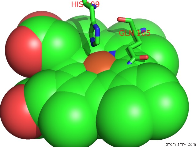
Mono view
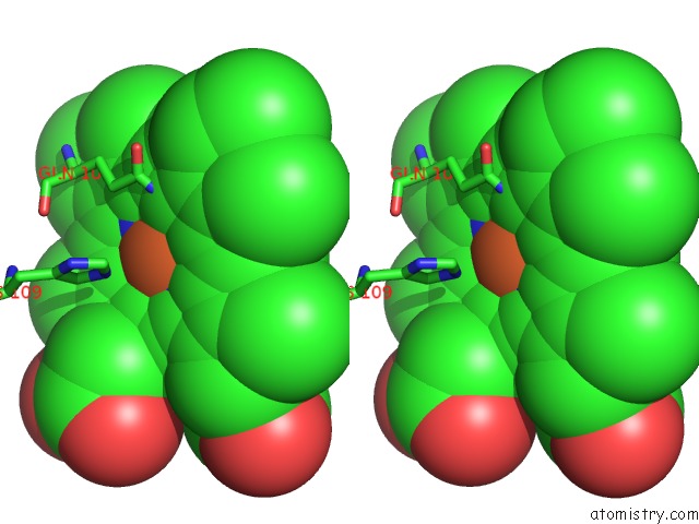
Stereo pair view
Mono view
Stereo pair view
A full contact list of Iron with other atoms in the Fe binding
site number 1 of First Structural Evidence of Substrate Specificity in Mammalian Peroxidases: Crystal Structures of Substrate Complexes with Lactoperoxidases From Two Different Species within 5.0Å range:
|
Reference:
I.A.Sheikh,
A.K.Singh,
N.Singh,
M.Sinha,
S.B.Singh,
A.Bhushan,
P.Kaur,
A.Srinivasan,
S.Sharma,
T.P.Singh.
Structural Evidence of Substrate Specificity in Mammalian Peroxidases: Structure of the Thiocyanate Complex with Lactoperoxidase and Its Interactions at 2.4 A Resolution J.Biol.Chem. V. 284 14849 2009.
ISSN: ISSN 0021-9258
PubMed: 19339248
DOI: 10.1074/JBC.M807644200
Page generated: Tue Aug 5 01:03:31 2025
ISSN: ISSN 0021-9258
PubMed: 19339248
DOI: 10.1074/JBC.M807644200
Last articles
Fe in 7BHCFe in 7BIU
Fe in 7BI1
Fe in 7B9A
Fe in 7BGI
Fe in 7BHB
Fe in 7B97
Fe in 7BHA
Fe in 7BBT
Fe in 7BET