Iron »
PDB 3ebd-3esf »
3esf »
Iron in PDB 3esf: Crystal Structure of the Enzyme Fe-Superoxide Dismutase TBSODB2 From Trypanosoma Brucei
Enzymatic activity of Crystal Structure of the Enzyme Fe-Superoxide Dismutase TBSODB2 From Trypanosoma Brucei
All present enzymatic activity of Crystal Structure of the Enzyme Fe-Superoxide Dismutase TBSODB2 From Trypanosoma Brucei:
1.15.1.1;
1.15.1.1;
Protein crystallography data
The structure of Crystal Structure of the Enzyme Fe-Superoxide Dismutase TBSODB2 From Trypanosoma Brucei, PDB code: 3esf
was solved by
J.F.R.Bachega,
M.V.A.S.Navarro,
R.C.Garratt,
with X-Ray Crystallography technique. A brief refinement statistics is given in the table below:
| Resolution Low / High (Å) | 30.18 / 2.01 |
| Space group | P 1 21 1 |
| Cell size a, b, c (Å), α, β, γ (°) | 68.302, 76.504, 76.981, 90.00, 92.70, 90.00 |
| R / Rfree (%) | 17.5 / 21.9 |
Iron Binding Sites:
The binding sites of Iron atom in the Crystal Structure of the Enzyme Fe-Superoxide Dismutase TBSODB2 From Trypanosoma Brucei
(pdb code 3esf). This binding sites where shown within
5.0 Angstroms radius around Iron atom.
In total 4 binding sites of Iron where determined in the Crystal Structure of the Enzyme Fe-Superoxide Dismutase TBSODB2 From Trypanosoma Brucei, PDB code: 3esf:
Jump to Iron binding site number: 1; 2; 3; 4;
In total 4 binding sites of Iron where determined in the Crystal Structure of the Enzyme Fe-Superoxide Dismutase TBSODB2 From Trypanosoma Brucei, PDB code: 3esf:
Jump to Iron binding site number: 1; 2; 3; 4;
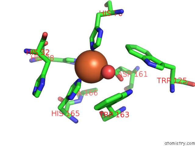
Iron binding site 1 out of 4 in 3esf
Go back to
Iron binding site 1 out
of 4 in the Crystal Structure of the Enzyme Fe-Superoxide Dismutase TBSODB2 From Trypanosoma Brucei
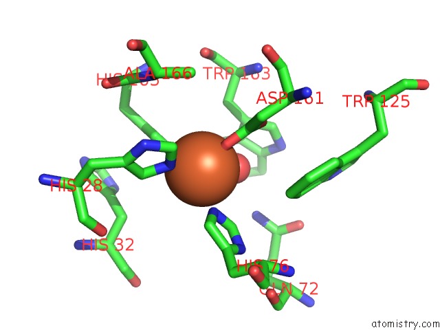
Mono view
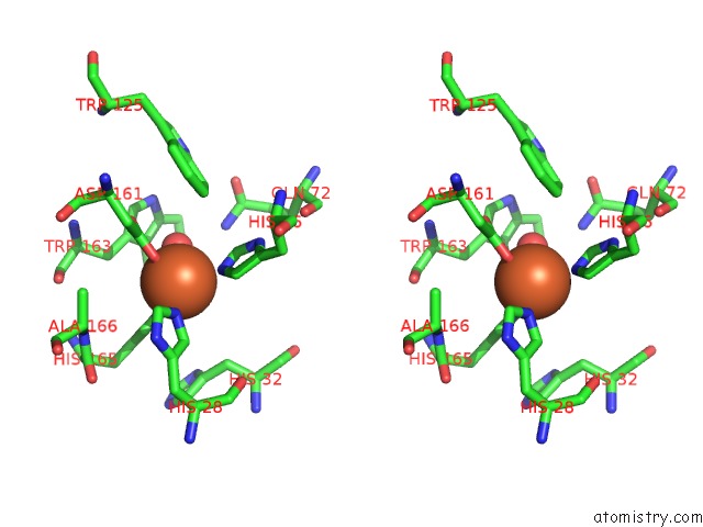
Stereo pair view
Mono view
Stereo pair view
A full contact list of Iron with other atoms in the Fe binding
site number 1 of Crystal Structure of the Enzyme Fe-Superoxide Dismutase TBSODB2 From Trypanosoma Brucei within 5.0Å range:
|
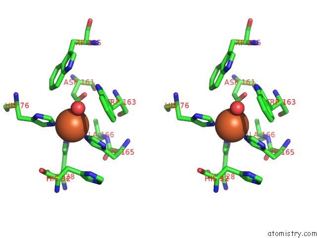
Iron binding site 2 out of 4 in 3esf
Go back to
Iron binding site 2 out
of 4 in the Crystal Structure of the Enzyme Fe-Superoxide Dismutase TBSODB2 From Trypanosoma Brucei
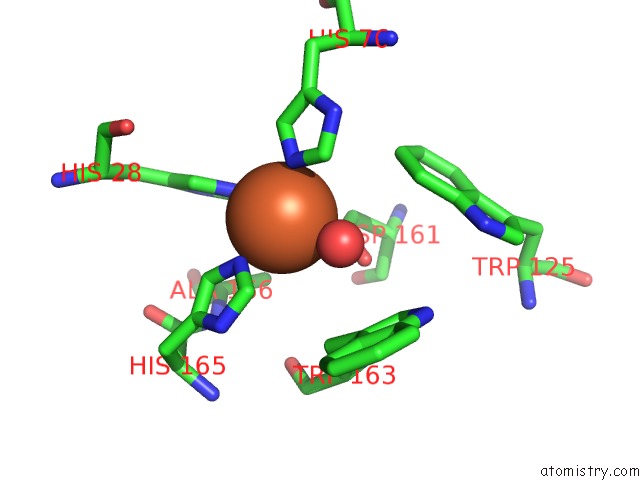
Mono view
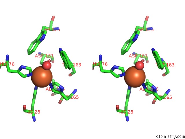
Stereo pair view
Mono view
Stereo pair view
A full contact list of Iron with other atoms in the Fe binding
site number 2 of Crystal Structure of the Enzyme Fe-Superoxide Dismutase TBSODB2 From Trypanosoma Brucei within 5.0Å range:
|
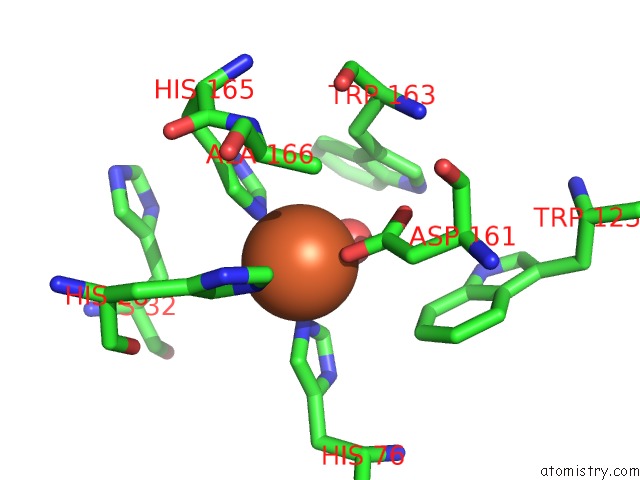
Iron binding site 3 out of 4 in 3esf
Go back to
Iron binding site 3 out
of 4 in the Crystal Structure of the Enzyme Fe-Superoxide Dismutase TBSODB2 From Trypanosoma Brucei
Mono view
Stereo pair view
Mono view
Stereo pair view
A full contact list of Iron with other atoms in the Fe binding
site number 3 of Crystal Structure of the Enzyme Fe-Superoxide Dismutase TBSODB2 From Trypanosoma Brucei within 5.0Å range:
|
Iron binding site 4 out of 4 in 3esf
Go back to
Iron binding site 4 out
of 4 in the Crystal Structure of the Enzyme Fe-Superoxide Dismutase TBSODB2 From Trypanosoma Brucei
Mono view
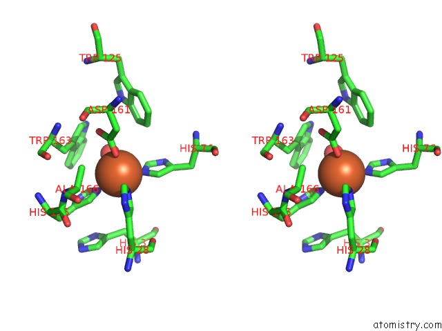
Stereo pair view
Mono view
Stereo pair view
A full contact list of Iron with other atoms in the Fe binding
site number 4 of Crystal Structure of the Enzyme Fe-Superoxide Dismutase TBSODB2 From Trypanosoma Brucei within 5.0Å range:
|
Reference:
J.F.Bachega,
M.V.Navarro,
L.Bleicher,
R.K.Bortoleto-Bugs,
D.Dive,
P.Hoffmann,
E.Viscogliosi,
R.C.Garratt.
Systematic Structural Studies of Iron Superoxide Dismutases From Human Parasites and A Statistical Coupling Analysis of Metal Binding Specificity Proteins V. 77 26 2009.
ISSN: ISSN 0887-3585
PubMed: 19384994
DOI: 10.1002/PROT.22412
Page generated: Tue Aug 5 01:03:48 2025
ISSN: ISSN 0887-3585
PubMed: 19384994
DOI: 10.1002/PROT.22412
Last articles
Yb in 1K90Yb in 1NCG
Yb in 1HS6
Yb in 1H19
Yb in 1C5K
Yb in 1GW6
Yb in 1CNT
Y in 8PUN
Y in 6PRF
Y in 8U7C