Iron »
PDB 3fpv-3gcj »
3fpw »
Iron in PDB 3fpw: Crystal Structure of Hbps with Bound Iron
Protein crystallography data
The structure of Crystal Structure of Hbps with Bound Iron, PDB code: 3fpw
was solved by
D.Ortiz De Orue Lucana,
G.Bogel,
P.Zou,
M.R.Groves,
with X-Ray Crystallography technique. A brief refinement statistics is given in the table below:
| Resolution Low / High (Å) | 18.80 / 1.60 |
| Space group | I 4 2 2 |
| Cell size a, b, c (Å), α, β, γ (°) | 77.940, 77.940, 79.990, 90.00, 90.00, 90.00 |
| R / Rfree (%) | 15.4 / 18.9 |
Iron Binding Sites:
The binding sites of Iron atom in the Crystal Structure of Hbps with Bound Iron
(pdb code 3fpw). This binding sites where shown within
5.0 Angstroms radius around Iron atom.
In total 2 binding sites of Iron where determined in the Crystal Structure of Hbps with Bound Iron, PDB code: 3fpw:
Jump to Iron binding site number: 1; 2;
In total 2 binding sites of Iron where determined in the Crystal Structure of Hbps with Bound Iron, PDB code: 3fpw:
Jump to Iron binding site number: 1; 2;
Iron binding site 1 out of 2 in 3fpw
Go back to
Iron binding site 1 out
of 2 in the Crystal Structure of Hbps with Bound Iron
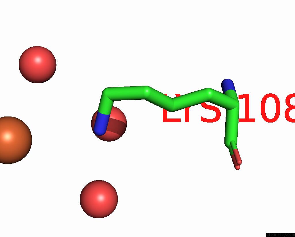
Mono view
Stereo pair view
Mono view
Stereo pair view
A full contact list of Iron with other atoms in the Fe binding
site number 1 of Crystal Structure of Hbps with Bound Iron within 5.0Å range:
|
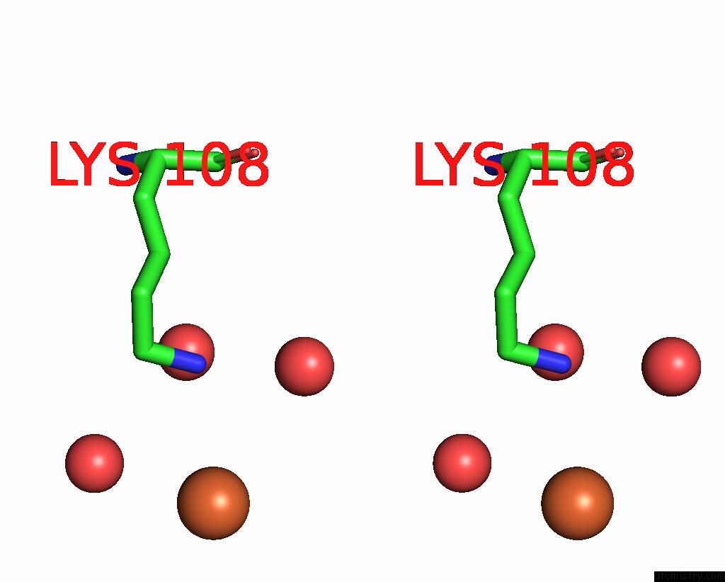
Iron binding site 2 out of 2 in 3fpw
Go back to
Iron binding site 2 out
of 2 in the Crystal Structure of Hbps with Bound Iron
Mono view
Stereo pair view
Mono view
Stereo pair view
A full contact list of Iron with other atoms in the Fe binding
site number 2 of Crystal Structure of Hbps with Bound Iron within 5.0Å range:
|
Reference:
D.Ortiz De Orue Lucana,
G.Bogel,
P.Zou,
M.R.Groves.
The Oligomeric Assembly of the Novel Haem-Degrading Protein Hbps Is Essential For Interaction with Its Cognate Two-Component Sensor Kinase J.Mol.Biol. V. 386 1108 2009.
ISSN: ISSN 0022-2836
PubMed: 19244623
DOI: 10.1016/J.JMB.2009.01.017
Page generated: Tue Aug 5 01:17:43 2025
ISSN: ISSN 0022-2836
PubMed: 19244623
DOI: 10.1016/J.JMB.2009.01.017
Last articles
Na in 6TBFNa in 6TBH
Na in 6TBG
Na in 6TB7
Na in 6TB1
Na in 6T99
Na in 6TA7
Na in 6T96
Na in 6T8A
Na in 6T89