Iron »
PDB 3fpv-3gcj »
3fvz »
Iron in PDB 3fvz: Structure of Peptidyl-Alpha-Hydroxyglycine Alpha-Amidating Lyase (Pal)
Enzymatic activity of Structure of Peptidyl-Alpha-Hydroxyglycine Alpha-Amidating Lyase (Pal)
All present enzymatic activity of Structure of Peptidyl-Alpha-Hydroxyglycine Alpha-Amidating Lyase (Pal):
4.3.2.5;
4.3.2.5;
Protein crystallography data
The structure of Structure of Peptidyl-Alpha-Hydroxyglycine Alpha-Amidating Lyase (Pal), PDB code: 3fvz
was solved by
E.E.Chufan,
M.De,
B.A.Eipper,
R.E.Mains,
L.M.Amzel,
with X-Ray Crystallography technique. A brief refinement statistics is given in the table below:
| Resolution Low / High (Å) | 39.22 / 2.35 |
| Space group | P 21 21 21 |
| Cell size a, b, c (Å), α, β, γ (°) | 52.176, 75.056, 97.474, 90.00, 90.00, 90.00 |
| R / Rfree (%) | 20.3 / 26.3 |
Other elements in 3fvz:
The structure of Structure of Peptidyl-Alpha-Hydroxyglycine Alpha-Amidating Lyase (Pal) also contains other interesting chemical elements:
| Calcium | (Ca) | 1 atom |
| Zinc | (Zn) | 1 atom |
Iron Binding Sites:
The binding sites of Iron atom in the Structure of Peptidyl-Alpha-Hydroxyglycine Alpha-Amidating Lyase (Pal)
(pdb code 3fvz). This binding sites where shown within
5.0 Angstroms radius around Iron atom.
In total only one binding site of Iron was determined in the Structure of Peptidyl-Alpha-Hydroxyglycine Alpha-Amidating Lyase (Pal), PDB code: 3fvz:
In total only one binding site of Iron was determined in the Structure of Peptidyl-Alpha-Hydroxyglycine Alpha-Amidating Lyase (Pal), PDB code: 3fvz:
Iron binding site 1 out of 1 in 3fvz
Go back to
Iron binding site 1 out
of 1 in the Structure of Peptidyl-Alpha-Hydroxyglycine Alpha-Amidating Lyase (Pal)
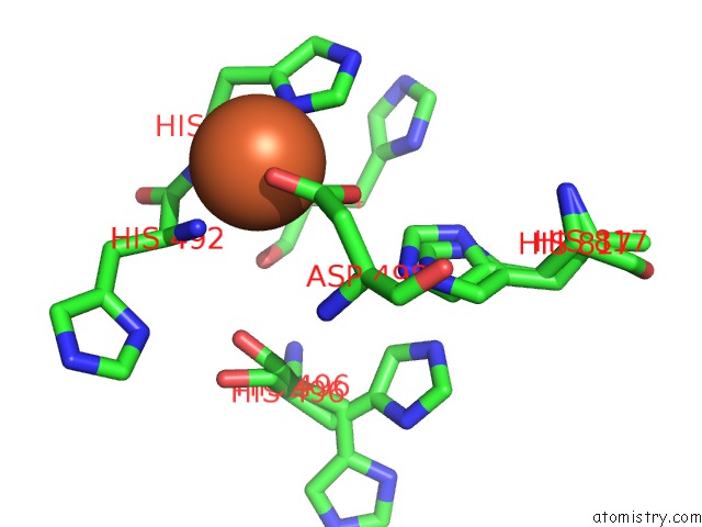
Mono view
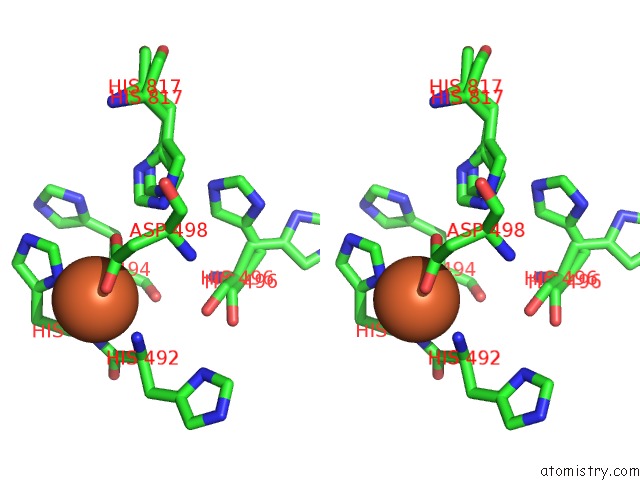
Stereo pair view
Mono view
Stereo pair view
A full contact list of Iron with other atoms in the Fe binding
site number 1 of Structure of Peptidyl-Alpha-Hydroxyglycine Alpha-Amidating Lyase (Pal) within 5.0Å range:
|
Reference:
E.E.Chufan,
M.De,
B.A.Eipper,
R.E.Mains,
L.M.Amzel.
Amidation of Bioactive Peptides: the Structure of the Lyase Domain of the Amidating Enzyme. Structure V. 17 965 2009.
ISSN: ISSN 0969-2126
PubMed: 19604476
DOI: 10.1016/J.STR.2009.05.008
Page generated: Tue Aug 5 01:18:08 2025
ISSN: ISSN 0969-2126
PubMed: 19604476
DOI: 10.1016/J.STR.2009.05.008
Last articles
Na in 5HXUNa in 5HXC
Na in 5HXI
Na in 5HWY
Na in 5HXA
Na in 5HWX
Na in 5HVD
Na in 5HVX
Na in 5HUT
Na in 5HTC