Iron »
PDB 3fpv-3gcj »
3fwy »
Iron in PDB 3fwy: Crystal Structure of the L Protein of Rhodobacter Sphaeroides Light- Independent Protochlorophyllide Reductase (Bchl) with Mgadp Bound: A Homologue of the Nitrogenase Fe Protein
Protein crystallography data
The structure of Crystal Structure of the L Protein of Rhodobacter Sphaeroides Light- Independent Protochlorophyllide Reductase (Bchl) with Mgadp Bound: A Homologue of the Nitrogenase Fe Protein, PDB code: 3fwy
was solved by
R.Sarma,
B.M.Barney,
T.L.Hamilton,
A.Jones,
L.C.Seefeldt,
J.W.Peters,
with X-Ray Crystallography technique. A brief refinement statistics is given in the table below:
| Resolution Low / High (Å) | 33.02 / 1.63 |
| Space group | P 21 21 21 |
| Cell size a, b, c (Å), α, β, γ (°) | 56.729, 86.622, 117.169, 90.00, 90.00, 90.00 |
| R / Rfree (%) | 17.4 / 19.9 |
Other elements in 3fwy:
The structure of Crystal Structure of the L Protein of Rhodobacter Sphaeroides Light- Independent Protochlorophyllide Reductase (Bchl) with Mgadp Bound: A Homologue of the Nitrogenase Fe Protein also contains other interesting chemical elements:
| Magnesium | (Mg) | 2 atoms |
Iron Binding Sites:
The binding sites of Iron atom in the Crystal Structure of the L Protein of Rhodobacter Sphaeroides Light- Independent Protochlorophyllide Reductase (Bchl) with Mgadp Bound: A Homologue of the Nitrogenase Fe Protein
(pdb code 3fwy). This binding sites where shown within
5.0 Angstroms radius around Iron atom.
In total 4 binding sites of Iron where determined in the Crystal Structure of the L Protein of Rhodobacter Sphaeroides Light- Independent Protochlorophyllide Reductase (Bchl) with Mgadp Bound: A Homologue of the Nitrogenase Fe Protein, PDB code: 3fwy:
Jump to Iron binding site number: 1; 2; 3; 4;
In total 4 binding sites of Iron where determined in the Crystal Structure of the L Protein of Rhodobacter Sphaeroides Light- Independent Protochlorophyllide Reductase (Bchl) with Mgadp Bound: A Homologue of the Nitrogenase Fe Protein, PDB code: 3fwy:
Jump to Iron binding site number: 1; 2; 3; 4;
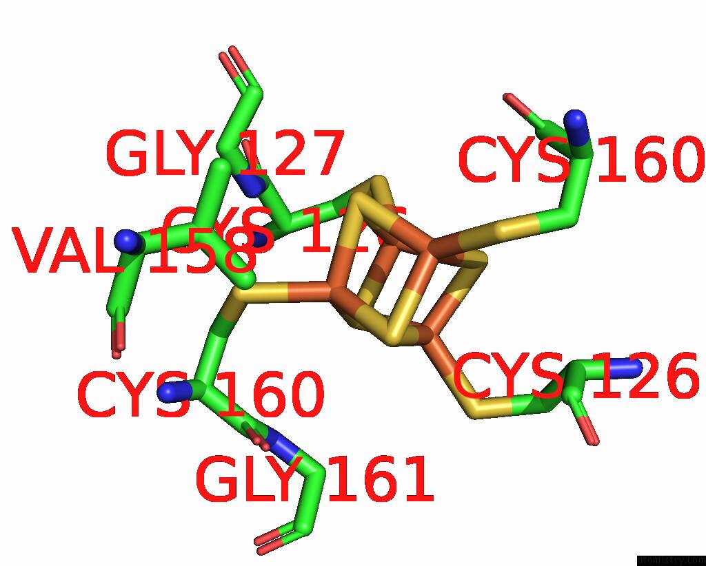
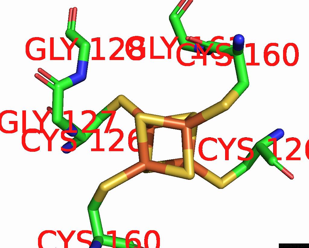
Iron binding site 1 out of 4 in 3fwy
Go back to
Iron binding site 1 out
of 4 in the Crystal Structure of the L Protein of Rhodobacter Sphaeroides Light- Independent Protochlorophyllide Reductase (Bchl) with Mgadp Bound: A Homologue of the Nitrogenase Fe Protein
Mono view
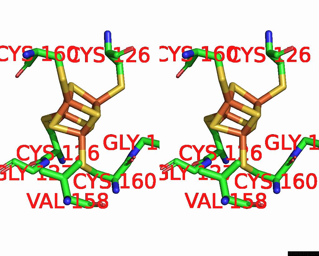
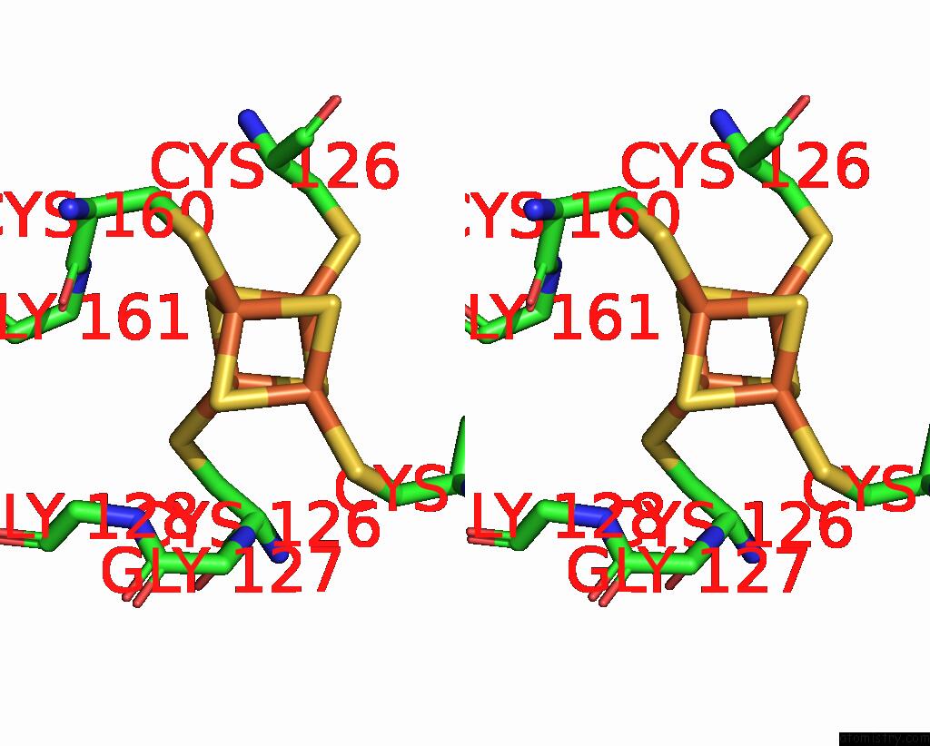
Stereo pair view
Mono view
Stereo pair view
A full contact list of Iron with other atoms in the Fe binding
site number 1 of Crystal Structure of the L Protein of Rhodobacter Sphaeroides Light- Independent Protochlorophyllide Reductase (Bchl) with Mgadp Bound: A Homologue of the Nitrogenase Fe Protein within 5.0Å range:
|
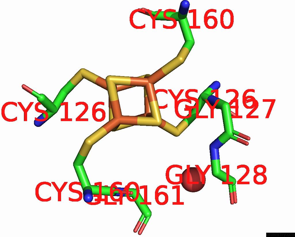
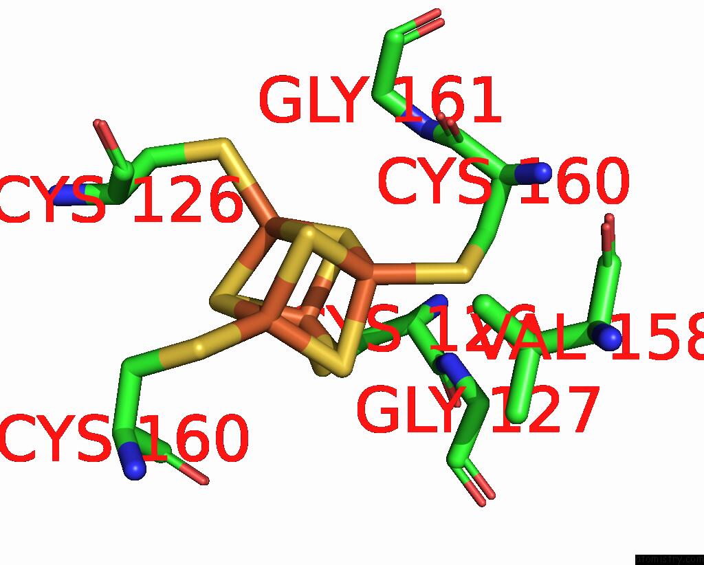
Iron binding site 2 out of 4 in 3fwy
Go back to
Iron binding site 2 out
of 4 in the Crystal Structure of the L Protein of Rhodobacter Sphaeroides Light- Independent Protochlorophyllide Reductase (Bchl) with Mgadp Bound: A Homologue of the Nitrogenase Fe Protein
Mono view
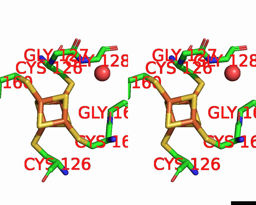
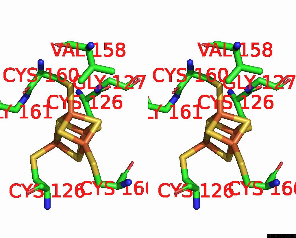
Stereo pair view
Mono view
Stereo pair view
A full contact list of Iron with other atoms in the Fe binding
site number 2 of Crystal Structure of the L Protein of Rhodobacter Sphaeroides Light- Independent Protochlorophyllide Reductase (Bchl) with Mgadp Bound: A Homologue of the Nitrogenase Fe Protein within 5.0Å range:
|
Iron binding site 3 out of 4 in 3fwy
Go back to
Iron binding site 3 out
of 4 in the Crystal Structure of the L Protein of Rhodobacter Sphaeroides Light- Independent Protochlorophyllide Reductase (Bchl) with Mgadp Bound: A Homologue of the Nitrogenase Fe Protein
Mono view
Stereo pair view
Mono view
Stereo pair view
A full contact list of Iron with other atoms in the Fe binding
site number 3 of Crystal Structure of the L Protein of Rhodobacter Sphaeroides Light- Independent Protochlorophyllide Reductase (Bchl) with Mgadp Bound: A Homologue of the Nitrogenase Fe Protein within 5.0Å range:
|
Iron binding site 4 out of 4 in 3fwy
Go back to
Iron binding site 4 out
of 4 in the Crystal Structure of the L Protein of Rhodobacter Sphaeroides Light- Independent Protochlorophyllide Reductase (Bchl) with Mgadp Bound: A Homologue of the Nitrogenase Fe Protein
Mono view
Stereo pair view
Mono view
Stereo pair view
A full contact list of Iron with other atoms in the Fe binding
site number 4 of Crystal Structure of the L Protein of Rhodobacter Sphaeroides Light- Independent Protochlorophyllide Reductase (Bchl) with Mgadp Bound: A Homologue of the Nitrogenase Fe Protein within 5.0Å range:
|
Reference:
R.Sarma,
B.M.Barney,
T.L.Hamilton,
A.Jones,
L.C.Seefeldt,
J.W.Peters.
Crystal Structure of the L Protein of Rhodobacter Sphaeroides Light-Independent Protochlorophyllide Reductase with Mgadp Bound: A Homologue of the Nitrogenase Fe Protein. Biochemistry V. 47 13004 2008.
ISSN: ISSN 0006-2960
PubMed: 19006326
DOI: 10.1021/BI801058R
Page generated: Sun Aug 4 10:20:39 2024
ISSN: ISSN 0006-2960
PubMed: 19006326
DOI: 10.1021/BI801058R
Last articles
Zn in 9MJ5Zn in 9HNW
Zn in 9G0L
Zn in 9FNE
Zn in 9DZN
Zn in 9E0I
Zn in 9D32
Zn in 9DAK
Zn in 8ZXC
Zn in 8ZUF