Iron »
PDB 3gck-3h33 »
3git »
Iron in PDB 3git: Crystal Structure of A Truncated Acetyl-Coa Synthase
Enzymatic activity of Crystal Structure of A Truncated Acetyl-Coa Synthase
All present enzymatic activity of Crystal Structure of A Truncated Acetyl-Coa Synthase:
2.3.1.169;
2.3.1.169;
Protein crystallography data
The structure of Crystal Structure of A Truncated Acetyl-Coa Synthase, PDB code: 3git
was solved by
A.Volbeda,
C.Darnault,
J.C.Fontecilla-Camps,
with X-Ray Crystallography technique. A brief refinement statistics is given in the table below:
| Resolution Low / High (Å) | 19.98 / 3.00 |
| Space group | P 31 2 1 |
| Cell size a, b, c (Å), α, β, γ (°) | 166.400, 166.400, 245.200, 90.00, 90.00, 120.00 |
| R / Rfree (%) | 17.1 / 20.8 |
Other elements in 3git:
The structure of Crystal Structure of A Truncated Acetyl-Coa Synthase also contains other interesting chemical elements:
| Zinc | (Zn) | 6 atoms |
Iron Binding Sites:
Pages:
>>> Page 1 <<< Page 2, Binding sites: 11 - 20; Page 3, Binding sites: 21 - 24;Binding sites:
The binding sites of Iron atom in the Crystal Structure of A Truncated Acetyl-Coa Synthase (pdb code 3git). This binding sites where shown within 5.0 Angstroms radius around Iron atom.In total 24 binding sites of Iron where determined in the Crystal Structure of A Truncated Acetyl-Coa Synthase, PDB code: 3git:
Jump to Iron binding site number: 1; 2; 3; 4; 5; 6; 7; 8; 9; 10;
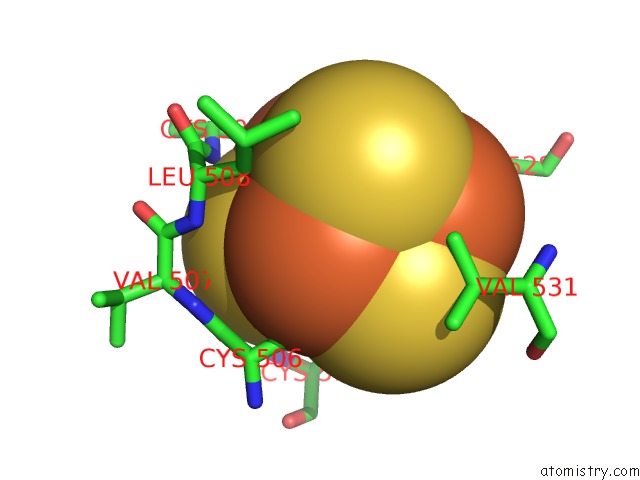
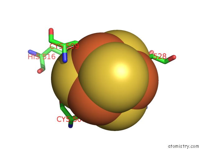
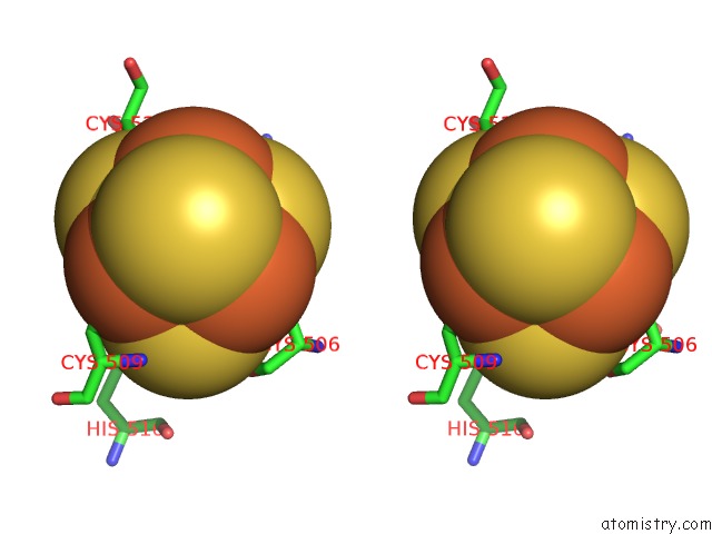
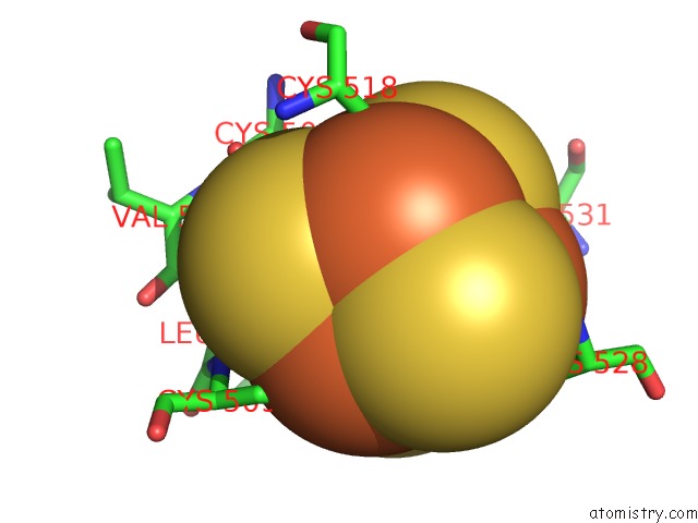
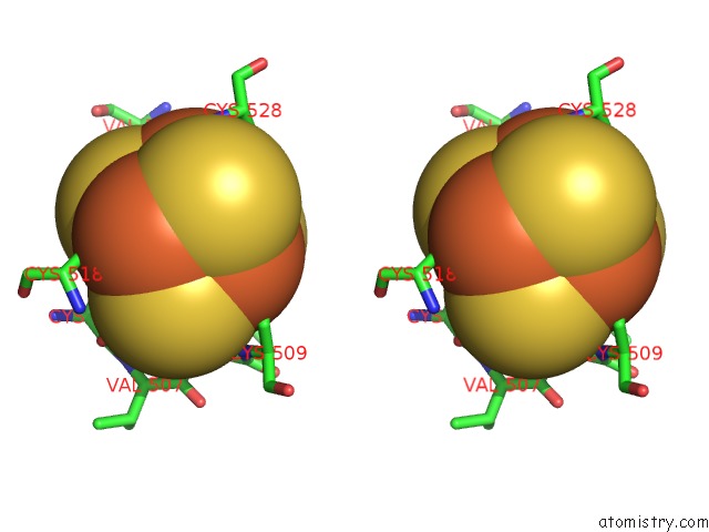
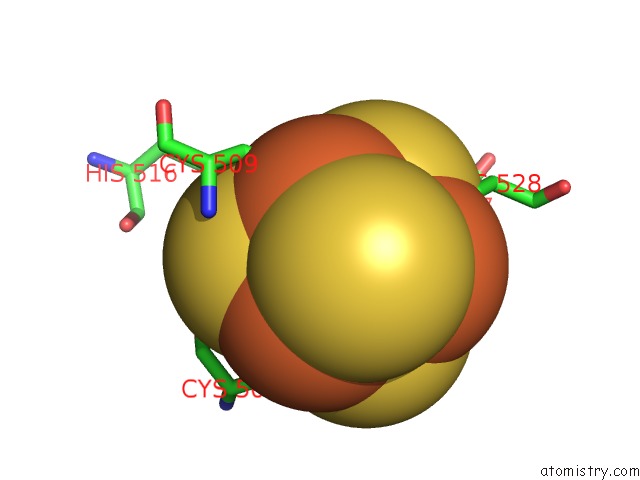
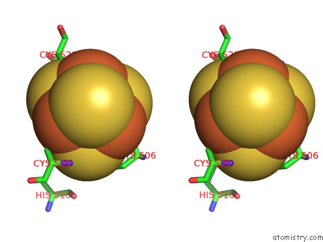
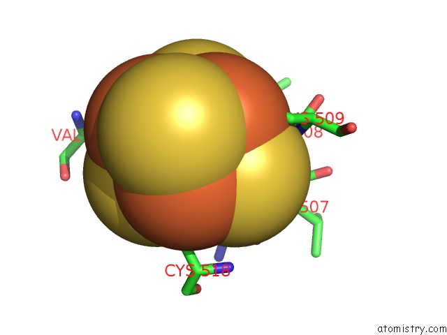
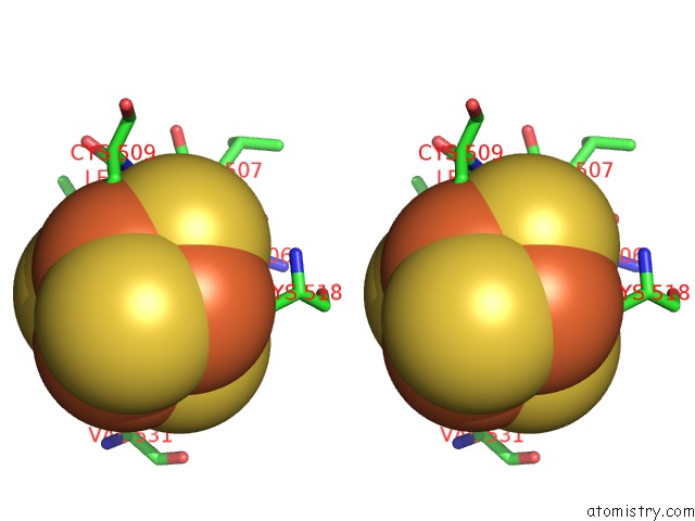
Iron binding site 1 out of 24 in 3git
Go back to
Iron binding site 1 out
of 24 in the Crystal Structure of A Truncated Acetyl-Coa Synthase
Mono view
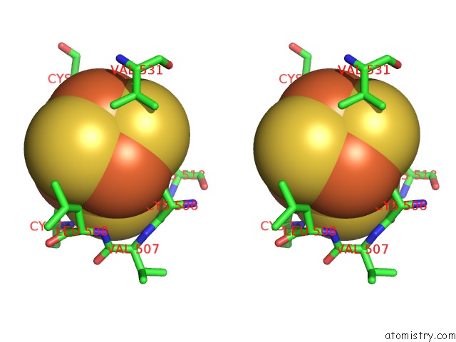
Stereo pair view
Mono view
Stereo pair view
|
|
A full contact list of Iron with other atoms in the Fe binding
site number 1 of Crystal Structure of A Truncated Acetyl-Coa Synthase within 5.0Å range:
|
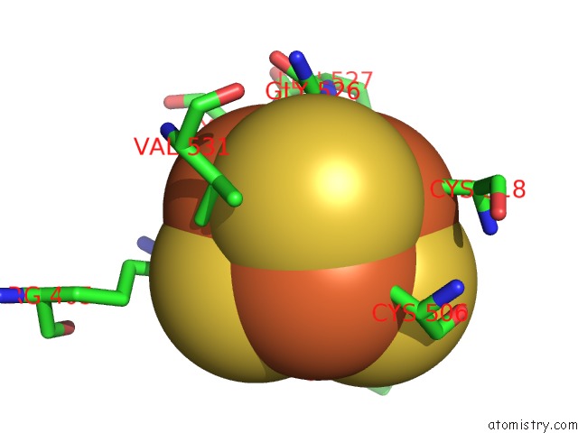
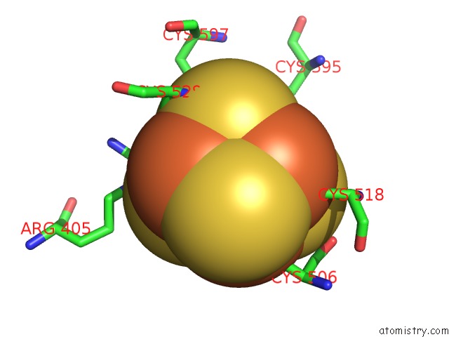
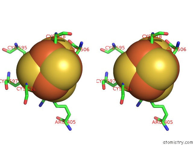
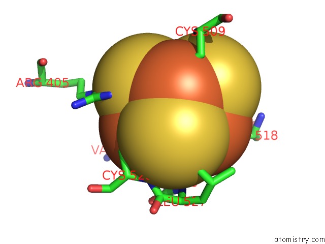
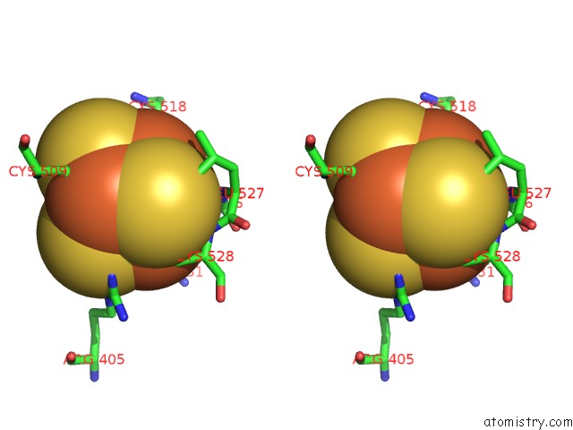
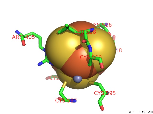
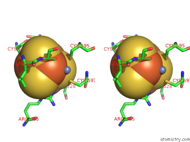
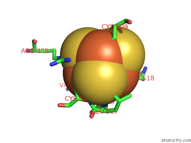
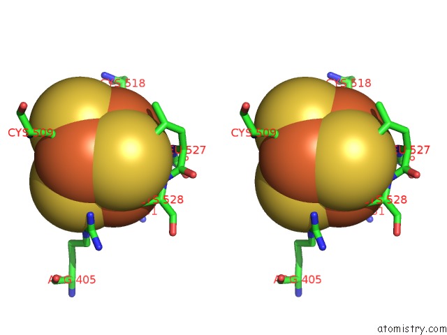
Iron binding site 2 out of 24 in 3git
Go back to
Iron binding site 2 out
of 24 in the Crystal Structure of A Truncated Acetyl-Coa Synthase
Mono view
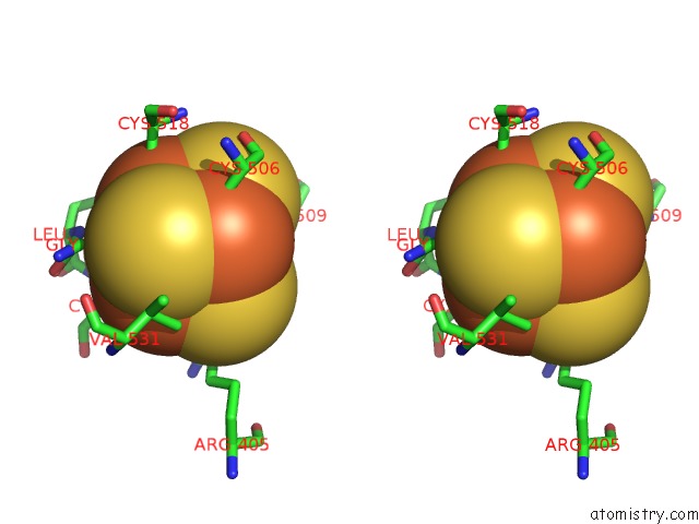
Stereo pair view
Mono view
Stereo pair view
|
|
A full contact list of Iron with other atoms in the Fe binding
site number 2 of Crystal Structure of A Truncated Acetyl-Coa Synthase within 5.0Å range:
|
Iron binding site 3 out of 24 in 3git
Go back to
Iron binding site 3 out
of 24 in the Crystal Structure of A Truncated Acetyl-Coa Synthase
Mono view
Stereo pair view
Mono view
Stereo pair view
|
|
A full contact list of Iron with other atoms in the Fe binding
site number 3 of Crystal Structure of A Truncated Acetyl-Coa Synthase within 5.0Å range:
|
Iron binding site 4 out of 24 in 3git
Go back to
Iron binding site 4 out
of 24 in the Crystal Structure of A Truncated Acetyl-Coa Synthase
Mono view
Stereo pair view
Mono view
Stereo pair view
|
|
A full contact list of Iron with other atoms in the Fe binding
site number 4 of Crystal Structure of A Truncated Acetyl-Coa Synthase within 5.0Å range:
|
Iron binding site 5 out of 24 in 3git
Go back to
Iron binding site 5 out
of 24 in the Crystal Structure of A Truncated Acetyl-Coa Synthase
Mono view
Stereo pair view
Mono view
Stereo pair view
|
|
A full contact list of Iron with other atoms in the Fe binding
site number 5 of Crystal Structure of A Truncated Acetyl-Coa Synthase within 5.0Å range:
|
Iron binding site 6 out of 24 in 3git
Go back to
Iron binding site 6 out
of 24 in the Crystal Structure of A Truncated Acetyl-Coa Synthase
Mono view
Stereo pair view
Mono view
Stereo pair view
|
|
A full contact list of Iron with other atoms in the Fe binding
site number 6 of Crystal Structure of A Truncated Acetyl-Coa Synthase within 5.0Å range:
|
Iron binding site 7 out of 24 in 3git
Go back to
Iron binding site 7 out
of 24 in the Crystal Structure of A Truncated Acetyl-Coa Synthase
Mono view
Stereo pair view
Mono view
Stereo pair view
|
|
A full contact list of Iron with other atoms in the Fe binding
site number 7 of Crystal Structure of A Truncated Acetyl-Coa Synthase within 5.0Å range:
|
Iron binding site 8 out of 24 in 3git
Go back to
Iron binding site 8 out
of 24 in the Crystal Structure of A Truncated Acetyl-Coa Synthase
Mono view
Stereo pair view
Mono view
Stereo pair view
|
|
A full contact list of Iron with other atoms in the Fe binding
site number 8 of Crystal Structure of A Truncated Acetyl-Coa Synthase within 5.0Å range:
|
Iron binding site 9 out of 24 in 3git
Go back to
Iron binding site 9 out
of 24 in the Crystal Structure of A Truncated Acetyl-Coa Synthase
Mono view
Stereo pair view
Mono view
Stereo pair view
|
|
A full contact list of Iron with other atoms in the Fe binding
site number 9 of Crystal Structure of A Truncated Acetyl-Coa Synthase within 5.0Å range:
|
Iron binding site 10 out of 24 in 3git
Go back to
Iron binding site 10 out
of 24 in the Crystal Structure of A Truncated Acetyl-Coa Synthase
Mono view
Stereo pair view
Mono view
Stereo pair view
|
|
A full contact list of Iron with other atoms in the Fe binding
site number 10 of Crystal Structure of A Truncated Acetyl-Coa Synthase within 5.0Å range:
|
Reference:
A.Volbeda,
C.Darnault,
X.Tan,
P.A.Lindahl,
J.C.Fontecilla-Camps.
Novel Domain Arrangement in the Crystal Structure of A Truncated Acetyl-Coa Synthase From Moorella Thermoacetica Biochemistry V. 48 7916 2009.
ISSN: ISSN 0006-2960
PubMed: 19650626
DOI: 10.1021/BI9003952
Page generated: Tue Aug 5 01:29:31 2025
ISSN: ISSN 0006-2960
PubMed: 19650626
DOI: 10.1021/BI9003952
Last articles
Mg in 7Y1WMg in 7Y1N
Mg in 7Y1M
Mg in 7Y1L
Mg in 7Y1K
Mg in 7Y1J
Mg in 7Y1H
Mg in 7XZR
Mg in 7XY9
Mg in 7XYB