Iron »
PDB 3hnj-3i8r »
3i0a »
Iron in PDB 3i0a: Crystal Structure of Siderocalin (Ngal, Lipocalin 2) K134A Mutant Complexed with Ferric Enterobactin
Protein crystallography data
The structure of Crystal Structure of Siderocalin (Ngal, Lipocalin 2) K134A Mutant Complexed with Ferric Enterobactin, PDB code: 3i0a
was solved by
M.C.Clifton,
with X-Ray Crystallography technique. A brief refinement statistics is given in the table below:
| Resolution Low / High (Å) | 50.00 / 2.60 |
| Space group | P 41 21 2 |
| Cell size a, b, c (Å), α, β, γ (°) | 114.253, 114.253, 117.950, 90.00, 90.00, 90.00 |
| R / Rfree (%) | 25.5 / 30 |
Iron Binding Sites:
The binding sites of Iron atom in the Crystal Structure of Siderocalin (Ngal, Lipocalin 2) K134A Mutant Complexed with Ferric Enterobactin
(pdb code 3i0a). This binding sites where shown within
5.0 Angstroms radius around Iron atom.
In total 3 binding sites of Iron where determined in the Crystal Structure of Siderocalin (Ngal, Lipocalin 2) K134A Mutant Complexed with Ferric Enterobactin, PDB code: 3i0a:
Jump to Iron binding site number: 1; 2; 3;
In total 3 binding sites of Iron where determined in the Crystal Structure of Siderocalin (Ngal, Lipocalin 2) K134A Mutant Complexed with Ferric Enterobactin, PDB code: 3i0a:
Jump to Iron binding site number: 1; 2; 3;
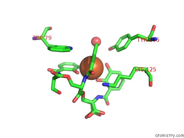
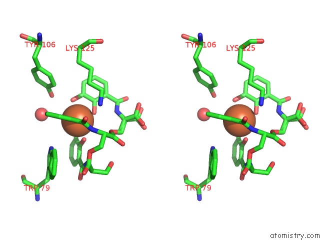
Iron binding site 1 out of 3 in 3i0a
Go back to
Iron binding site 1 out
of 3 in the Crystal Structure of Siderocalin (Ngal, Lipocalin 2) K134A Mutant Complexed with Ferric Enterobactin
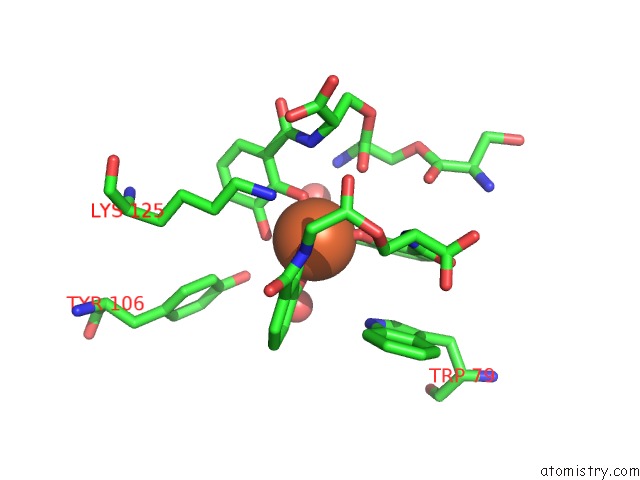
Mono view
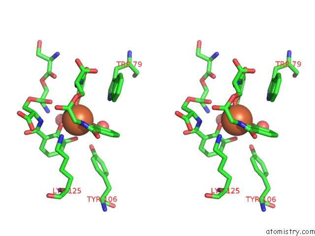
Stereo pair view
Mono view
Stereo pair view
A full contact list of Iron with other atoms in the Fe binding
site number 1 of Crystal Structure of Siderocalin (Ngal, Lipocalin 2) K134A Mutant Complexed with Ferric Enterobactin within 5.0Å range:
|
Iron binding site 2 out of 3 in 3i0a
Go back to
Iron binding site 2 out
of 3 in the Crystal Structure of Siderocalin (Ngal, Lipocalin 2) K134A Mutant Complexed with Ferric Enterobactin
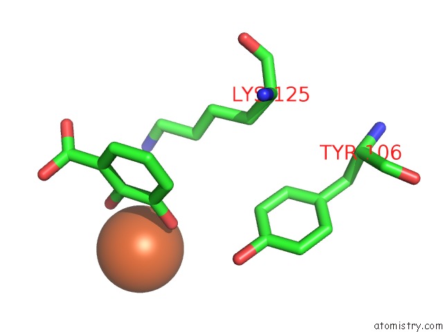
Mono view
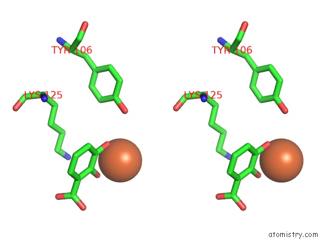
Stereo pair view
Mono view
Stereo pair view
A full contact list of Iron with other atoms in the Fe binding
site number 2 of Crystal Structure of Siderocalin (Ngal, Lipocalin 2) K134A Mutant Complexed with Ferric Enterobactin within 5.0Å range:
|
Iron binding site 3 out of 3 in 3i0a
Go back to
Iron binding site 3 out
of 3 in the Crystal Structure of Siderocalin (Ngal, Lipocalin 2) K134A Mutant Complexed with Ferric Enterobactin
Mono view
Stereo pair view
Mono view
Stereo pair view
A full contact list of Iron with other atoms in the Fe binding
site number 3 of Crystal Structure of Siderocalin (Ngal, Lipocalin 2) K134A Mutant Complexed with Ferric Enterobactin within 5.0Å range:
|
Reference:
M.C.Clifton,
P.B.Rupert,
T.M.Hoette,
K.N.Raymond,
R.J.Abergel,
R.K.Strong.
Parsing the Functional Specificity of Siderocalin / Lipocalin 2 / Ngal For Siderophores and Related Small-Molecule Ligands To Be Published.
Page generated: Tue Aug 5 02:08:13 2025
Last articles
Na in 3T0DNa in 3T0B
Na in 3T2J
Na in 3T2I
Na in 3T2H
Na in 3T09
Na in 3SZS
Na in 3T08
Na in 3SZ9
Na in 3SZT