Iron »
PDB 3k9z-3kyw »
3kn7 »
Iron in PDB 3kn7: Crystal Structure of Haemophilus Influenzae Y195A Mutant Holo Ferric Ion-Binding Protein A
Protein crystallography data
The structure of Crystal Structure of Haemophilus Influenzae Y195A Mutant Holo Ferric Ion-Binding Protein A, PDB code: 3kn7
was solved by
S.R.Shouldice,
A.B.Schryvers,
with X-Ray Crystallography technique. A brief refinement statistics is given in the table below:
| Resolution Low / High (Å) | 62.00 / 1.71 |
| Space group | P 21 21 2 |
| Cell size a, b, c (Å), α, β, γ (°) | 106.464, 75.822, 34.109, 90.00, 90.00, 90.00 |
| R / Rfree (%) | 19 / 23.7 |
Iron Binding Sites:
The binding sites of Iron atom in the Crystal Structure of Haemophilus Influenzae Y195A Mutant Holo Ferric Ion-Binding Protein A
(pdb code 3kn7). This binding sites where shown within
5.0 Angstroms radius around Iron atom.
In total only one binding site of Iron was determined in the Crystal Structure of Haemophilus Influenzae Y195A Mutant Holo Ferric Ion-Binding Protein A, PDB code: 3kn7:
In total only one binding site of Iron was determined in the Crystal Structure of Haemophilus Influenzae Y195A Mutant Holo Ferric Ion-Binding Protein A, PDB code: 3kn7:
Iron binding site 1 out of 1 in 3kn7
Go back to
Iron binding site 1 out
of 1 in the Crystal Structure of Haemophilus Influenzae Y195A Mutant Holo Ferric Ion-Binding Protein A
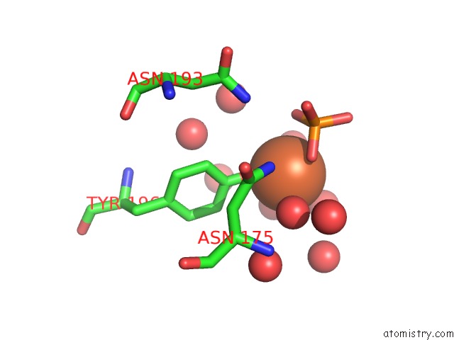
Mono view
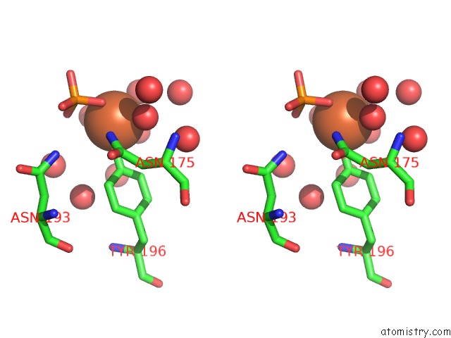
Stereo pair view
Mono view
Stereo pair view
A full contact list of Iron with other atoms in the Fe binding
site number 1 of Crystal Structure of Haemophilus Influenzae Y195A Mutant Holo Ferric Ion-Binding Protein A within 5.0Å range:
|
Reference:
H.K.Khambati,
T.F.Moraes,
J.Singh,
S.R.Shouldice,
R.H.Yu,
A.B.Schryvers.
The Role of Vicinal Tyrosine Residues in the Function of Haemophilus Influenzae Ferric Binding Protein A. Biochem.J. 2010.
ISSN: ESSN 1470-8728
PubMed: 20799927
DOI: 10.1042/BJ20101043
Page generated: Sun Aug 4 13:56:00 2024
ISSN: ESSN 1470-8728
PubMed: 20799927
DOI: 10.1042/BJ20101043
Last articles
Br in 1UV5Br in 1US0
Br in 1UPJ
Br in 1UHJ
Br in 1UHX
Br in 1UHY
Br in 1SR4
Br in 1TO3
Br in 1THC
Br in 1T0S