Iron »
PDB 3k9z-3kyw »
3ks5 »
Iron in PDB 3ks5: Crystal Structure of Putative Glycerophosphoryl Diester Phosphodiesterase (17743486) From Agrobacterium Tumefaciens Str. C58 (Dupont) at 2.05 A Resolution
Protein crystallography data
The structure of Crystal Structure of Putative Glycerophosphoryl Diester Phosphodiesterase (17743486) From Agrobacterium Tumefaciens Str. C58 (Dupont) at 2.05 A Resolution, PDB code: 3ks5
was solved by
Joint Center For Structural Genomics (Jcsg),
with X-Ray Crystallography technique. A brief refinement statistics is given in the table below:
| Resolution Low / High (Å) | 30.07 / 2.05 |
| Space group | P 41 21 2 |
| Cell size a, b, c (Å), α, β, γ (°) | 84.153, 84.153, 214.882, 90.00, 90.00, 90.00 |
| R / Rfree (%) | 21.2 / 24.4 |
Iron Binding Sites:
The binding sites of Iron atom in the Crystal Structure of Putative Glycerophosphoryl Diester Phosphodiesterase (17743486) From Agrobacterium Tumefaciens Str. C58 (Dupont) at 2.05 A Resolution
(pdb code 3ks5). This binding sites where shown within
5.0 Angstroms radius around Iron atom.
In total 2 binding sites of Iron where determined in the Crystal Structure of Putative Glycerophosphoryl Diester Phosphodiesterase (17743486) From Agrobacterium Tumefaciens Str. C58 (Dupont) at 2.05 A Resolution, PDB code: 3ks5:
Jump to Iron binding site number: 1; 2;
In total 2 binding sites of Iron where determined in the Crystal Structure of Putative Glycerophosphoryl Diester Phosphodiesterase (17743486) From Agrobacterium Tumefaciens Str. C58 (Dupont) at 2.05 A Resolution, PDB code: 3ks5:
Jump to Iron binding site number: 1; 2;
Iron binding site 1 out of 2 in 3ks5
Go back to
Iron binding site 1 out
of 2 in the Crystal Structure of Putative Glycerophosphoryl Diester Phosphodiesterase (17743486) From Agrobacterium Tumefaciens Str. C58 (Dupont) at 2.05 A Resolution
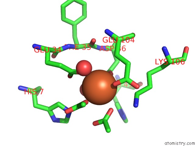
Mono view
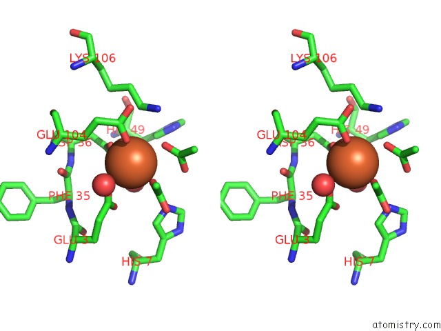
Stereo pair view
Mono view
Stereo pair view
A full contact list of Iron with other atoms in the Fe binding
site number 1 of Crystal Structure of Putative Glycerophosphoryl Diester Phosphodiesterase (17743486) From Agrobacterium Tumefaciens Str. C58 (Dupont) at 2.05 A Resolution within 5.0Å range:
|
Iron binding site 2 out of 2 in 3ks5
Go back to
Iron binding site 2 out
of 2 in the Crystal Structure of Putative Glycerophosphoryl Diester Phosphodiesterase (17743486) From Agrobacterium Tumefaciens Str. C58 (Dupont) at 2.05 A Resolution
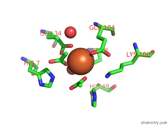
Mono view
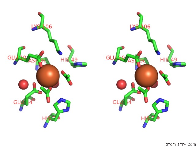
Stereo pair view
Mono view
Stereo pair view
A full contact list of Iron with other atoms in the Fe binding
site number 2 of Crystal Structure of Putative Glycerophosphoryl Diester Phosphodiesterase (17743486) From Agrobacterium Tumefaciens Str. C58 (Dupont) at 2.05 A Resolution within 5.0Å range:
|
Reference:
Joint Center For Structural Genomics (Jcsg),
Joint Center For Structural Genomics (Jcsg).
N/A N/A.
Page generated: Sun Aug 4 13:58:50 2024
Last articles
Br in 2FCCBr in 2FCY
Br in 2FCX
Br in 2EWF
Br in 2FCV
Br in 2F5M
Br in 2EZ0
Br in 2F4G
Br in 2D6B
Br in 2E4I