Iron »
PDB 3kyx-3lhb »
3l4p »
Iron in PDB 3l4p: Crystal Structure of the Aldehyde Dehydrogenase (A.K.A. Aor or Mop) of Desulfovibrio Gigas Covalently Bound to [ASO3]-
Enzymatic activity of Crystal Structure of the Aldehyde Dehydrogenase (A.K.A. Aor or Mop) of Desulfovibrio Gigas Covalently Bound to [ASO3]-
All present enzymatic activity of Crystal Structure of the Aldehyde Dehydrogenase (A.K.A. Aor or Mop) of Desulfovibrio Gigas Covalently Bound to [ASO3]-:
1.2.99.7;
1.2.99.7;
Protein crystallography data
The structure of Crystal Structure of the Aldehyde Dehydrogenase (A.K.A. Aor or Mop) of Desulfovibrio Gigas Covalently Bound to [ASO3]-, PDB code: 3l4p
was solved by
D.R.Boer,
M.J.Romao,
with X-Ray Crystallography technique. A brief refinement statistics is given in the table below:
| Resolution Low / High (Å) | 19.44 / 1.45 |
| Space group | P 61 2 2 |
| Cell size a, b, c (Å), α, β, γ (°) | 142.890, 142.890, 161.640, 90.00, 90.00, 120.00 |
| R / Rfree (%) | 14.2 / 16.6 |
Other elements in 3l4p:
The structure of Crystal Structure of the Aldehyde Dehydrogenase (A.K.A. Aor or Mop) of Desulfovibrio Gigas Covalently Bound to [ASO3]- also contains other interesting chemical elements:
| Molybdenum | (Mo) | 1 atom |
| Magnesium | (Mg) | 5 atoms |
| Arsenic | (As) | 1 atom |
| Calcium | (Ca) | 1 atom |
| Chlorine | (Cl) | 9 atoms |
Iron Binding Sites:
The binding sites of Iron atom in the Crystal Structure of the Aldehyde Dehydrogenase (A.K.A. Aor or Mop) of Desulfovibrio Gigas Covalently Bound to [ASO3]-
(pdb code 3l4p). This binding sites where shown within
5.0 Angstroms radius around Iron atom.
In total 4 binding sites of Iron where determined in the Crystal Structure of the Aldehyde Dehydrogenase (A.K.A. Aor or Mop) of Desulfovibrio Gigas Covalently Bound to [ASO3]-, PDB code: 3l4p:
Jump to Iron binding site number: 1; 2; 3; 4;
In total 4 binding sites of Iron where determined in the Crystal Structure of the Aldehyde Dehydrogenase (A.K.A. Aor or Mop) of Desulfovibrio Gigas Covalently Bound to [ASO3]-, PDB code: 3l4p:
Jump to Iron binding site number: 1; 2; 3; 4;
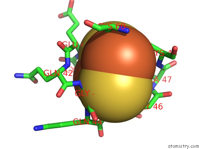
Iron binding site 1 out of 4 in 3l4p
Go back to
Iron binding site 1 out
of 4 in the Crystal Structure of the Aldehyde Dehydrogenase (A.K.A. Aor or Mop) of Desulfovibrio Gigas Covalently Bound to [ASO3]-
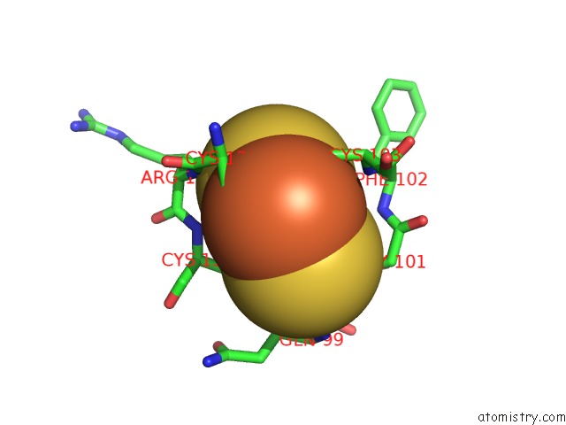
Mono view
Stereo pair view
Mono view
Stereo pair view
A full contact list of Iron with other atoms in the Fe binding
site number 1 of Crystal Structure of the Aldehyde Dehydrogenase (A.K.A. Aor or Mop) of Desulfovibrio Gigas Covalently Bound to [ASO3]- within 5.0Å range:
|
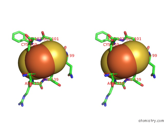
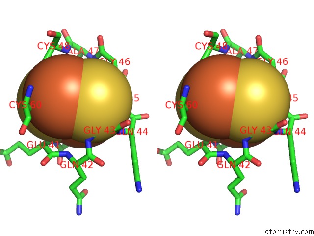
Iron binding site 2 out of 4 in 3l4p
Go back to
Iron binding site 2 out
of 4 in the Crystal Structure of the Aldehyde Dehydrogenase (A.K.A. Aor or Mop) of Desulfovibrio Gigas Covalently Bound to [ASO3]-
Mono view
Stereo pair view
Mono view
Stereo pair view
A full contact list of Iron with other atoms in the Fe binding
site number 2 of Crystal Structure of the Aldehyde Dehydrogenase (A.K.A. Aor or Mop) of Desulfovibrio Gigas Covalently Bound to [ASO3]- within 5.0Å range:
|
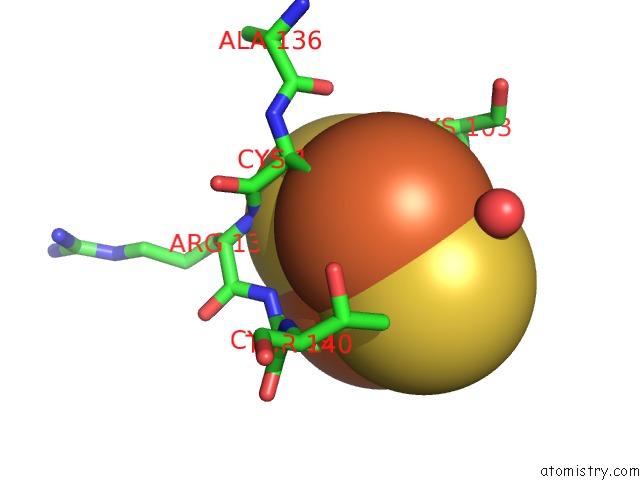
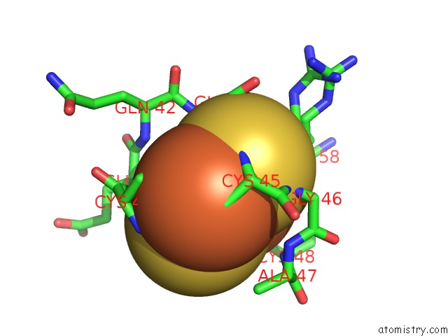
Iron binding site 3 out of 4 in 3l4p
Go back to
Iron binding site 3 out
of 4 in the Crystal Structure of the Aldehyde Dehydrogenase (A.K.A. Aor or Mop) of Desulfovibrio Gigas Covalently Bound to [ASO3]-
Mono view
Stereo pair view
Mono view
Stereo pair view
A full contact list of Iron with other atoms in the Fe binding
site number 3 of Crystal Structure of the Aldehyde Dehydrogenase (A.K.A. Aor or Mop) of Desulfovibrio Gigas Covalently Bound to [ASO3]- within 5.0Å range:
|
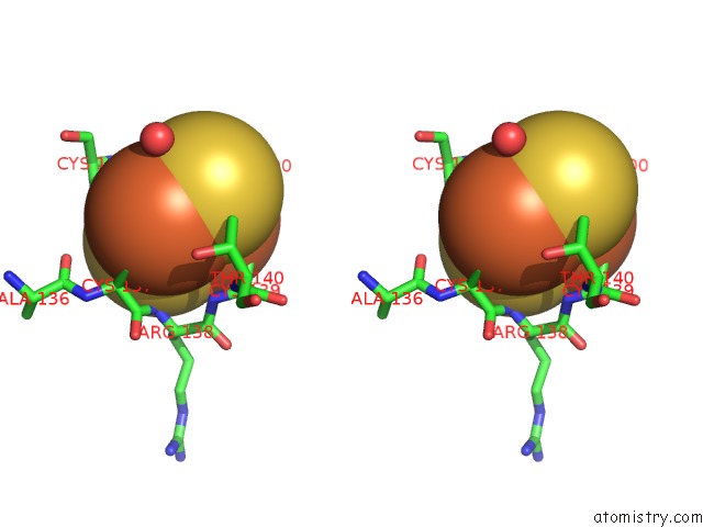
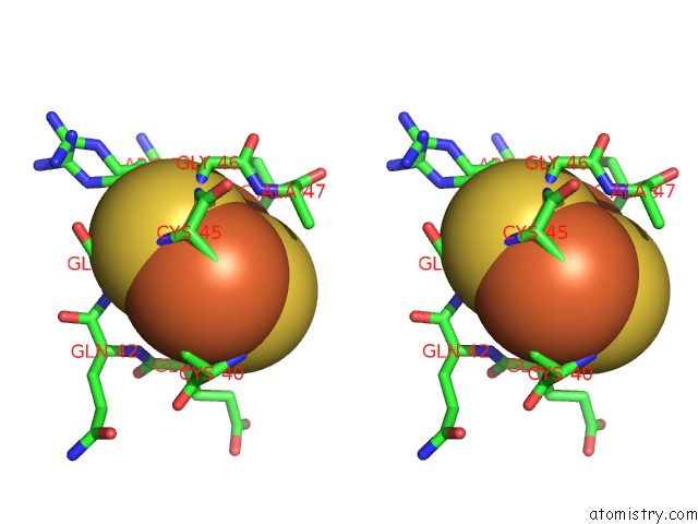
Iron binding site 4 out of 4 in 3l4p
Go back to
Iron binding site 4 out
of 4 in the Crystal Structure of the Aldehyde Dehydrogenase (A.K.A. Aor or Mop) of Desulfovibrio Gigas Covalently Bound to [ASO3]-
Mono view
Stereo pair view
Mono view
Stereo pair view
A full contact list of Iron with other atoms in the Fe binding
site number 4 of Crystal Structure of the Aldehyde Dehydrogenase (A.K.A. Aor or Mop) of Desulfovibrio Gigas Covalently Bound to [ASO3]- within 5.0Å range:
|
Reference:
A.Thapper,
D.R.Boer,
C.D.Brondino,
J.J.Moura,
M.J.Romao.
Correlating Epr and X-Ray Structural Analysis of Arsenite-Inhibited Forms of Aldehyde Oxidoreductase. J.Biol.Inorg.Chem. V. 12 353 2007.
ISSN: ISSN 0949-8257
PubMed: 17139522
DOI: 10.1007/S00775-006-0191-9
Page generated: Tue Aug 5 03:21:17 2025
ISSN: ISSN 0949-8257
PubMed: 17139522
DOI: 10.1007/S00775-006-0191-9
Last articles
Fe in 7VT2Fe in 7VXU
Fe in 7VY3
Fe in 7VXP
Fe in 7VXQ
Fe in 7VWJ
Fe in 7VW6
Fe in 7VX0
Fe in 7VVS
Fe in 7VW4