Iron »
PDB 3kyx-3lhb »
3l61 »
Iron in PDB 3l61: Crystal Structure of Substrate-Free P450CAM at 200 Mm [K+]
Enzymatic activity of Crystal Structure of Substrate-Free P450CAM at 200 Mm [K+]
All present enzymatic activity of Crystal Structure of Substrate-Free P450CAM at 200 Mm [K+]:
1.14.15.1;
1.14.15.1;
Protein crystallography data
The structure of Crystal Structure of Substrate-Free P450CAM at 200 Mm [K+], PDB code: 3l61
was solved by
Y.-T.Lee,
R.F.Wilson,
I.Rupniewski,
D.B.Goodin,
with X-Ray Crystallography technique. A brief refinement statistics is given in the table below:
| Resolution Low / High (Å) | 10.00 / 1.50 |
| Space group | P 21 21 21 |
| Cell size a, b, c (Å), α, β, γ (°) | 65.606, 73.825, 92.428, 90.00, 90.00, 90.00 |
| R / Rfree (%) | 19.7 / 21.6 |
Iron Binding Sites:
The binding sites of Iron atom in the Crystal Structure of Substrate-Free P450CAM at 200 Mm [K+]
(pdb code 3l61). This binding sites where shown within
5.0 Angstroms radius around Iron atom.
In total only one binding site of Iron was determined in the Crystal Structure of Substrate-Free P450CAM at 200 Mm [K+], PDB code: 3l61:
In total only one binding site of Iron was determined in the Crystal Structure of Substrate-Free P450CAM at 200 Mm [K+], PDB code: 3l61:
Iron binding site 1 out of 1 in 3l61
Go back to
Iron binding site 1 out
of 1 in the Crystal Structure of Substrate-Free P450CAM at 200 Mm [K+]
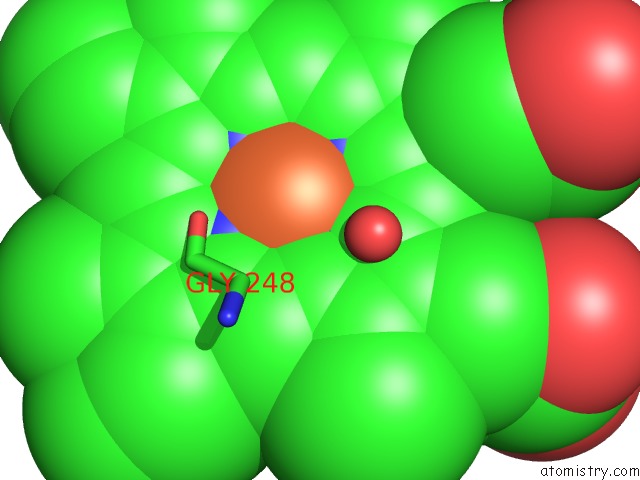
Mono view
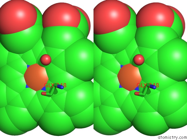
Stereo pair view
Mono view
Stereo pair view
A full contact list of Iron with other atoms in the Fe binding
site number 1 of Crystal Structure of Substrate-Free P450CAM at 200 Mm [K+] within 5.0Å range:
|
Reference:
Y.T.Lee,
R.F.Wilson,
I.Rupniewski,
D.B.Goodin.
P450CAM Visits An Open Conformation in the Absence of Substrate. Biochemistry V. 49 3412 2010.
ISSN: ISSN 0006-2960
PubMed: 20297780
DOI: 10.1021/BI100183G
Page generated: Tue Aug 5 03:21:25 2025
ISSN: ISSN 0006-2960
PubMed: 20297780
DOI: 10.1021/BI100183G
Last articles
Na in 8VMZNa in 8VMY
Na in 8VMX
Na in 8VMW
Na in 8VL8
Na in 8VMU
Na in 8VMT
Na in 8VMV
Na in 8VMS
Na in 8VMR