Iron »
PDB 3lhs-3m2i »
3lm4 »
Iron in PDB 3lm4: Crystal Structure of 2,3-Dihydroxy Biphenyl Dioxygenase From Rhodococcus Sp. (Strain RHA1)
Enzymatic activity of Crystal Structure of 2,3-Dihydroxy Biphenyl Dioxygenase From Rhodococcus Sp. (Strain RHA1)
All present enzymatic activity of Crystal Structure of 2,3-Dihydroxy Biphenyl Dioxygenase From Rhodococcus Sp. (Strain RHA1):
1.13.11.2;
1.13.11.2;
Protein crystallography data
The structure of Crystal Structure of 2,3-Dihydroxy Biphenyl Dioxygenase From Rhodococcus Sp. (Strain RHA1), PDB code: 3lm4
was solved by
B.Syed Ibrahim,
D.Kumaran,
S.K.Burley,
S.Swaminathan,
New Yorksgx Research Center For Structural Genomics (Nysgxrc),
with X-Ray Crystallography technique. A brief refinement statistics is given in the table below:
| Resolution Low / High (Å) | 37.67 / 1.80 |
| Space group | P 21 21 21 |
| Cell size a, b, c (Å), α, β, γ (°) | 74.923, 76.189, 251.373, 90.00, 90.00, 90.00 |
| R / Rfree (%) | 18.4 / 21 |
Iron Binding Sites:
The binding sites of Iron atom in the Crystal Structure of 2,3-Dihydroxy Biphenyl Dioxygenase From Rhodococcus Sp. (Strain RHA1)
(pdb code 3lm4). This binding sites where shown within
5.0 Angstroms radius around Iron atom.
In total 4 binding sites of Iron where determined in the Crystal Structure of 2,3-Dihydroxy Biphenyl Dioxygenase From Rhodococcus Sp. (Strain RHA1), PDB code: 3lm4:
Jump to Iron binding site number: 1; 2; 3; 4;
In total 4 binding sites of Iron where determined in the Crystal Structure of 2,3-Dihydroxy Biphenyl Dioxygenase From Rhodococcus Sp. (Strain RHA1), PDB code: 3lm4:
Jump to Iron binding site number: 1; 2; 3; 4;
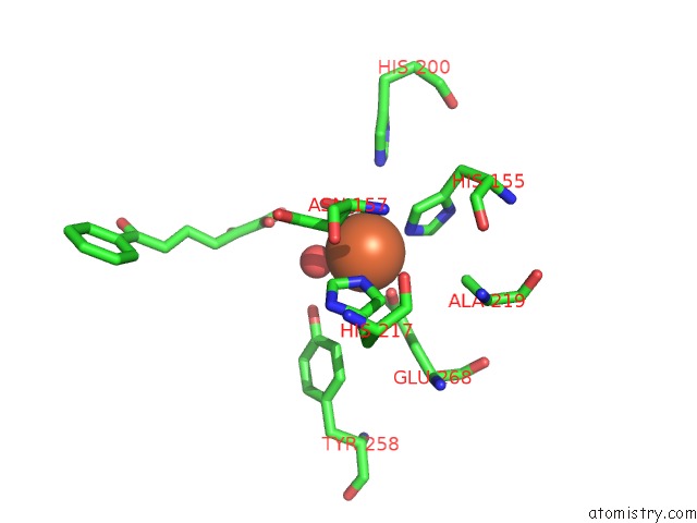
Iron binding site 1 out of 4 in 3lm4
Go back to
Iron binding site 1 out
of 4 in the Crystal Structure of 2,3-Dihydroxy Biphenyl Dioxygenase From Rhodococcus Sp. (Strain RHA1)
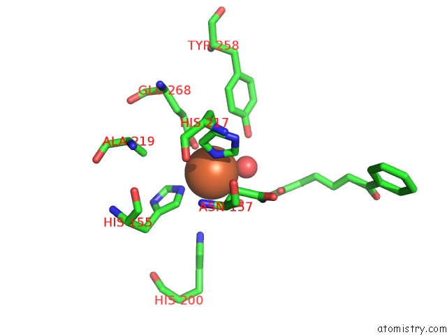
Mono view
Stereo pair view
Mono view
Stereo pair view
A full contact list of Iron with other atoms in the Fe binding
site number 1 of Crystal Structure of 2,3-Dihydroxy Biphenyl Dioxygenase From Rhodococcus Sp. (Strain RHA1) within 5.0Å range:
|
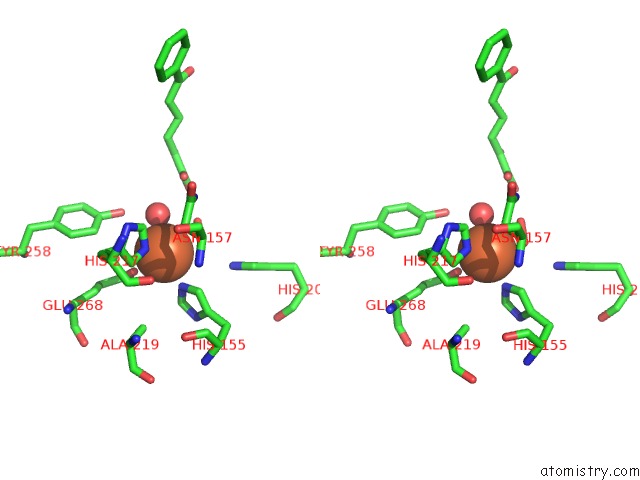
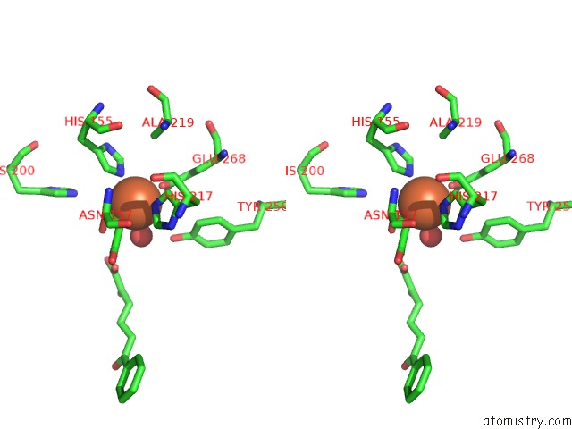
Iron binding site 2 out of 4 in 3lm4
Go back to
Iron binding site 2 out
of 4 in the Crystal Structure of 2,3-Dihydroxy Biphenyl Dioxygenase From Rhodococcus Sp. (Strain RHA1)
Mono view
Stereo pair view
Mono view
Stereo pair view
A full contact list of Iron with other atoms in the Fe binding
site number 2 of Crystal Structure of 2,3-Dihydroxy Biphenyl Dioxygenase From Rhodococcus Sp. (Strain RHA1) within 5.0Å range:
|
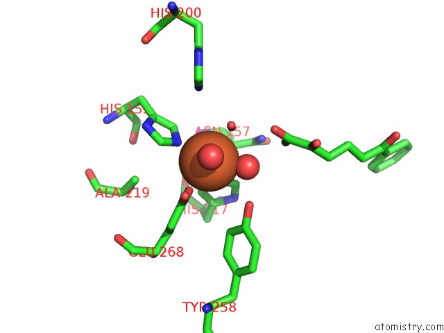
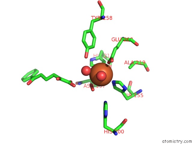
Iron binding site 3 out of 4 in 3lm4
Go back to
Iron binding site 3 out
of 4 in the Crystal Structure of 2,3-Dihydroxy Biphenyl Dioxygenase From Rhodococcus Sp. (Strain RHA1)
Mono view
Stereo pair view
Mono view
Stereo pair view
A full contact list of Iron with other atoms in the Fe binding
site number 3 of Crystal Structure of 2,3-Dihydroxy Biphenyl Dioxygenase From Rhodococcus Sp. (Strain RHA1) within 5.0Å range:
|
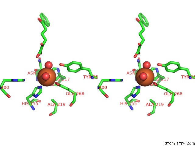
Iron binding site 4 out of 4 in 3lm4
Go back to
Iron binding site 4 out
of 4 in the Crystal Structure of 2,3-Dihydroxy Biphenyl Dioxygenase From Rhodococcus Sp. (Strain RHA1)
Mono view
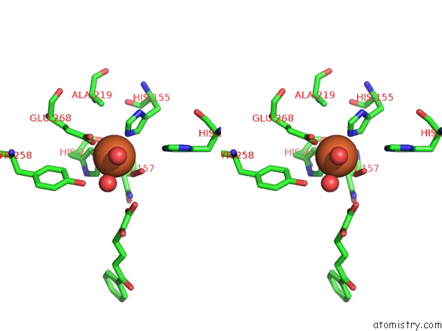
Stereo pair view
Mono view
Stereo pair view
A full contact list of Iron with other atoms in the Fe binding
site number 4 of Crystal Structure of 2,3-Dihydroxy Biphenyl Dioxygenase From Rhodococcus Sp. (Strain RHA1) within 5.0Å range:
|
Reference:
B.Syed Ibrahim,
D.Kumaran,
S.K.Burley,
S.Swaminathan.
Crystal Structure of 2,3-Dihydroxy Biphenyl Dioxygenase From Rhodococcus Sp. (Strain RHA1) To Be Published.
Page generated: Sun Aug 4 14:30:19 2024
Last articles
Fe in 2YXOFe in 2YRS
Fe in 2YXC
Fe in 2YNM
Fe in 2YVJ
Fe in 2YP1
Fe in 2YU2
Fe in 2YU1
Fe in 2YQB
Fe in 2YOO