Iron »
PDB 3m38-3mma »
3m97 »
Iron in PDB 3m97: Structure of the Soluble Domain of Cytochrome C552 with Its Flexible Linker Segment From Paracoccus Denitrificans
Protein crystallography data
The structure of Structure of the Soluble Domain of Cytochrome C552 with Its Flexible Linker Segment From Paracoccus Denitrificans, PDB code: 3m97
was solved by
C.Rajendran,
U.Ermler,
B.Ludwig,
H.Michel,
with X-Ray Crystallography technique. A brief refinement statistics is given in the table below:
| Resolution Low / High (Å) | 29.13 / 1.33 |
| Space group | P 21 21 21 |
| Cell size a, b, c (Å), α, β, γ (°) | 37.376, 38.991, 93.014, 90.00, 90.00, 90.00 |
| R / Rfree (%) | 20 / 22.9 |
Other elements in 3m97:
The structure of Structure of the Soluble Domain of Cytochrome C552 with Its Flexible Linker Segment From Paracoccus Denitrificans also contains other interesting chemical elements:
| Zinc | (Zn) | 4 atoms |
Iron Binding Sites:
The binding sites of Iron atom in the Structure of the Soluble Domain of Cytochrome C552 with Its Flexible Linker Segment From Paracoccus Denitrificans
(pdb code 3m97). This binding sites where shown within
5.0 Angstroms radius around Iron atom.
In total only one binding site of Iron was determined in the Structure of the Soluble Domain of Cytochrome C552 with Its Flexible Linker Segment From Paracoccus Denitrificans, PDB code: 3m97:
In total only one binding site of Iron was determined in the Structure of the Soluble Domain of Cytochrome C552 with Its Flexible Linker Segment From Paracoccus Denitrificans, PDB code: 3m97:
Iron binding site 1 out of 1 in 3m97
Go back to
Iron binding site 1 out
of 1 in the Structure of the Soluble Domain of Cytochrome C552 with Its Flexible Linker Segment From Paracoccus Denitrificans
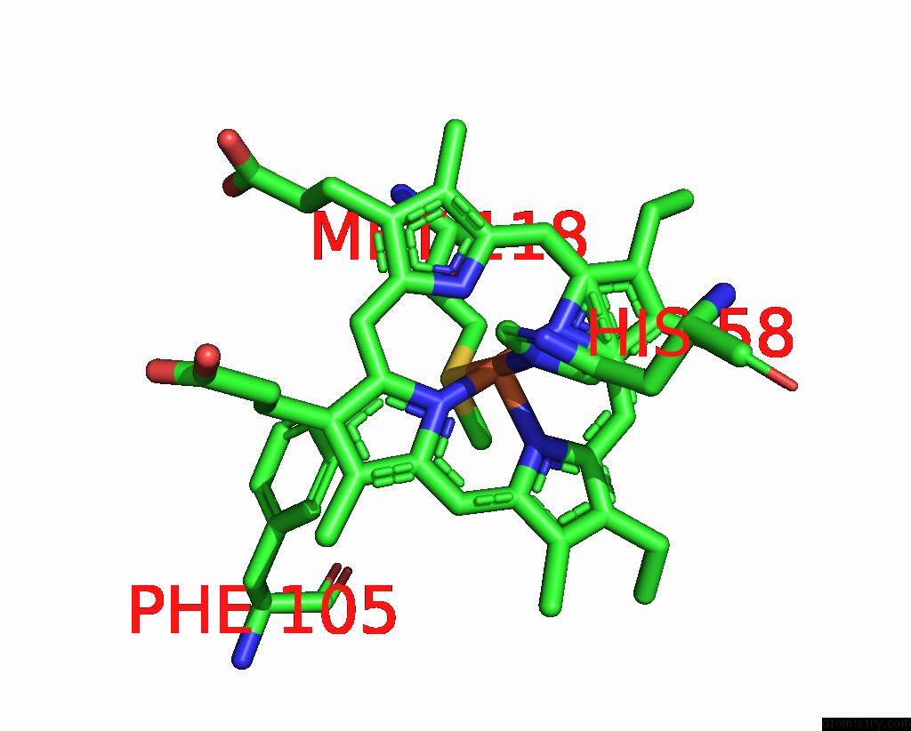
Mono view
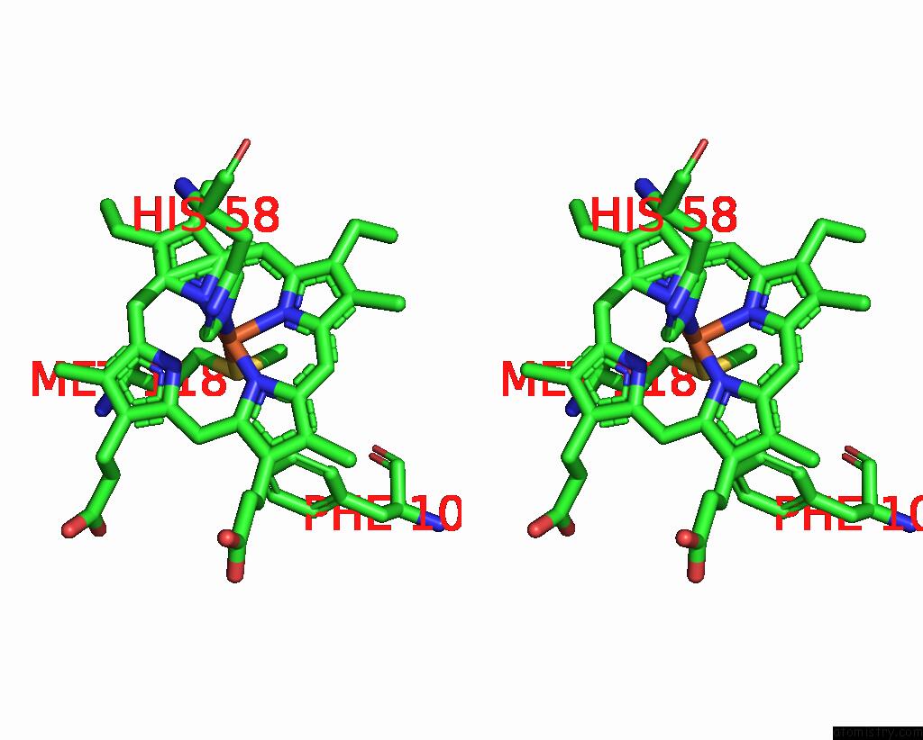
Stereo pair view
Mono view
Stereo pair view
A full contact list of Iron with other atoms in the Fe binding
site number 1 of Structure of the Soluble Domain of Cytochrome C552 with Its Flexible Linker Segment From Paracoccus Denitrificans within 5.0Å range:
|
Reference:
C.Rajendran,
U.Ermler,
B.Ludwig,
H.Michel.
Structure at 1.5 A Resolution of Cytochrome C(552) with Its Flexible Linker Segment, A Membrane-Anchored Protein From Paracoccus Denitrificans. Acta Crystallogr.,Sect.D V. 66 850 2010.
ISSN: ISSN 0907-4449
PubMed: 20606266
DOI: 10.1107/S0907444910019396
Page generated: Tue Aug 5 03:44:49 2025
ISSN: ISSN 0907-4449
PubMed: 20606266
DOI: 10.1107/S0907444910019396
Last articles
Fe in 5CNDFe in 5CNC
Fe in 5CNB
Fe in 5CN9
Fe in 5CN8
Fe in 5CN7
Fe in 5CN6
Fe in 5CN5
Fe in 5CN4
Fe in 5CJH