Iron »
PDB 3nxu-3ol5 »
3nxu »
Iron in PDB 3nxu: Crystal Structure of Human Cytochrome P4503A4 Bound to An Inhibitor Ritonavir
Enzymatic activity of Crystal Structure of Human Cytochrome P4503A4 Bound to An Inhibitor Ritonavir
All present enzymatic activity of Crystal Structure of Human Cytochrome P4503A4 Bound to An Inhibitor Ritonavir:
1.14.13.32; 1.14.13.67; 1.14.13.97;
1.14.13.32; 1.14.13.67; 1.14.13.97;
Protein crystallography data
The structure of Crystal Structure of Human Cytochrome P4503A4 Bound to An Inhibitor Ritonavir, PDB code: 3nxu
was solved by
I.F.Sevrioukova,
T.L.Poulos,
with X-Ray Crystallography technique. A brief refinement statistics is given in the table below:
| Resolution Low / High (Å) | 40.40 / 2.00 |
| Space group | C 1 2 1 |
| Cell size a, b, c (Å), α, β, γ (°) | 162.123, 94.690, 93.130, 90.00, 124.25, 90.00 |
| R / Rfree (%) | 23.2 / 26.2 |
Iron Binding Sites:
The binding sites of Iron atom in the Crystal Structure of Human Cytochrome P4503A4 Bound to An Inhibitor Ritonavir
(pdb code 3nxu). This binding sites where shown within
5.0 Angstroms radius around Iron atom.
In total 2 binding sites of Iron where determined in the Crystal Structure of Human Cytochrome P4503A4 Bound to An Inhibitor Ritonavir, PDB code: 3nxu:
Jump to Iron binding site number: 1; 2;
In total 2 binding sites of Iron where determined in the Crystal Structure of Human Cytochrome P4503A4 Bound to An Inhibitor Ritonavir, PDB code: 3nxu:
Jump to Iron binding site number: 1; 2;
Iron binding site 1 out of 2 in 3nxu
Go back to
Iron binding site 1 out
of 2 in the Crystal Structure of Human Cytochrome P4503A4 Bound to An Inhibitor Ritonavir
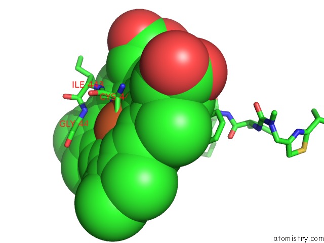
Mono view
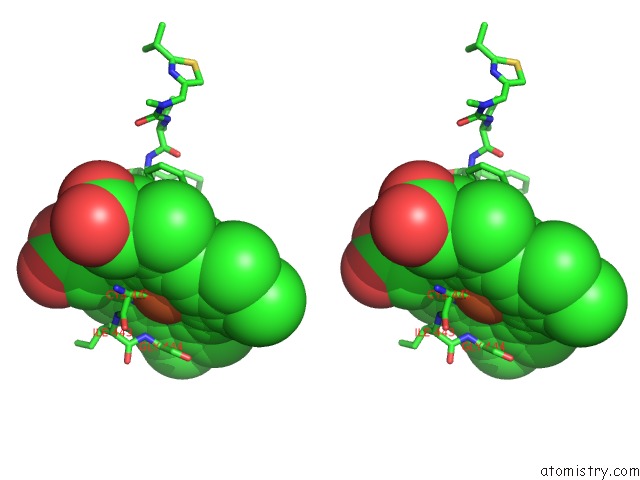
Stereo pair view
Mono view
Stereo pair view
A full contact list of Iron with other atoms in the Fe binding
site number 1 of Crystal Structure of Human Cytochrome P4503A4 Bound to An Inhibitor Ritonavir within 5.0Å range:
|
Iron binding site 2 out of 2 in 3nxu
Go back to
Iron binding site 2 out
of 2 in the Crystal Structure of Human Cytochrome P4503A4 Bound to An Inhibitor Ritonavir
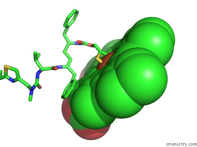
Mono view
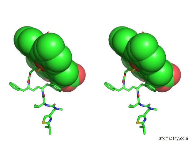
Stereo pair view
Mono view
Stereo pair view
A full contact list of Iron with other atoms in the Fe binding
site number 2 of Crystal Structure of Human Cytochrome P4503A4 Bound to An Inhibitor Ritonavir within 5.0Å range:
|
Reference:
I.F.Sevrioukova,
T.L.Poulos.
Structure and Mechanism of the Complex Between Cytochrome P4503A4 and Ritonavir. Proc.Natl.Acad.Sci.Usa V. 107 18422 2010.
ISSN: ISSN 0027-8424
PubMed: 20937904
DOI: 10.1073/PNAS.1010693107
Page generated: Tue Aug 5 05:02:08 2025
ISSN: ISSN 0027-8424
PubMed: 20937904
DOI: 10.1073/PNAS.1010693107
Last articles
Na in 4CCGNa in 4CBX
Na in 4C90
Na in 4C7A
Na in 4C80
Na in 4C79
Na in 4C76
Na in 4C6Y
Na in 4C6S
Na in 4C75