Iron »
PDB 3nxu-3ol5 »
3o1a »
Iron in PDB 3o1a: Structure of Oxye (CYP165D3), A Cytochrome P450 Involved in Teicoplanin Biosynthesis
Enzymatic activity of Structure of Oxye (CYP165D3), A Cytochrome P450 Involved in Teicoplanin Biosynthesis
All present enzymatic activity of Structure of Oxye (CYP165D3), A Cytochrome P450 Involved in Teicoplanin Biosynthesis:
1.14.14.1;
1.14.14.1;
Protein crystallography data
The structure of Structure of Oxye (CYP165D3), A Cytochrome P450 Involved in Teicoplanin Biosynthesis, PDB code: 3o1a
was solved by
M.J.Cryle,
I.Schlichting,
with X-Ray Crystallography technique. A brief refinement statistics is given in the table below:
| Resolution Low / High (Å) | 49.27 / 2.50 |
| Space group | P 31 |
| Cell size a, b, c (Å), α, β, γ (°) | 75.680, 75.680, 74.720, 90.00, 90.00, 120.00 |
| R / Rfree (%) | 22 / 27.4 |
Iron Binding Sites:
The binding sites of Iron atom in the Structure of Oxye (CYP165D3), A Cytochrome P450 Involved in Teicoplanin Biosynthesis
(pdb code 3o1a). This binding sites where shown within
5.0 Angstroms radius around Iron atom.
In total only one binding site of Iron was determined in the Structure of Oxye (CYP165D3), A Cytochrome P450 Involved in Teicoplanin Biosynthesis, PDB code: 3o1a:
In total only one binding site of Iron was determined in the Structure of Oxye (CYP165D3), A Cytochrome P450 Involved in Teicoplanin Biosynthesis, PDB code: 3o1a:
Iron binding site 1 out of 1 in 3o1a
Go back to
Iron binding site 1 out
of 1 in the Structure of Oxye (CYP165D3), A Cytochrome P450 Involved in Teicoplanin Biosynthesis
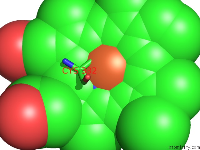
Mono view
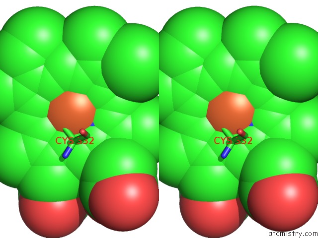
Stereo pair view
Mono view
Stereo pair view
A full contact list of Iron with other atoms in the Fe binding
site number 1 of Structure of Oxye (CYP165D3), A Cytochrome P450 Involved in Teicoplanin Biosynthesis within 5.0Å range:
|
Reference:
M.J.Cryle,
J.Staaden,
I.Schlichting.
Structural Characterization of CYP165D3, A Cytochrome P450 Involved in Phenolic Coupling in Teicoplanin Biosynthesis. Arch.Biochem.Biophys. V. 507 163 2011.
ISSN: ISSN 0003-9861
PubMed: 20974107
DOI: 10.1016/J.ABB.2010.10.017
Page generated: Tue Aug 5 05:02:07 2025
ISSN: ISSN 0003-9861
PubMed: 20974107
DOI: 10.1016/J.ABB.2010.10.017
Last articles
Na in 1H80Na in 1H18
Na in 1H6M
Na in 1H5V
Na in 1H17
Na in 1H16
Na in 1GZG
Na in 1GWD
Na in 1GZ1
Na in 1GWU