Iron »
PDB 3nxu-3ol5 »
3obz »
Iron in PDB 3obz: Crystal Structure of Human Phytanoyl-Coa Dioxygenase PHYHD1 2- Oxoglutarate and Iron Complex
Protein crystallography data
The structure of Crystal Structure of Human Phytanoyl-Coa Dioxygenase PHYHD1 2- Oxoglutarate and Iron Complex, PDB code: 3obz
was solved by
Z.Zhang,
M.A.Mcdonough,
C.J.Schofield,
with X-Ray Crystallography technique. A brief refinement statistics is given in the table below:
| Resolution Low / High (Å) | 24.31 / 2.15 |
| Space group | P 31 2 1 |
| Cell size a, b, c (Å), α, β, γ (°) | 92.366, 92.366, 81.754, 90.00, 90.00, 120.00 |
| R / Rfree (%) | 23.3 / 27.4 |
Iron Binding Sites:
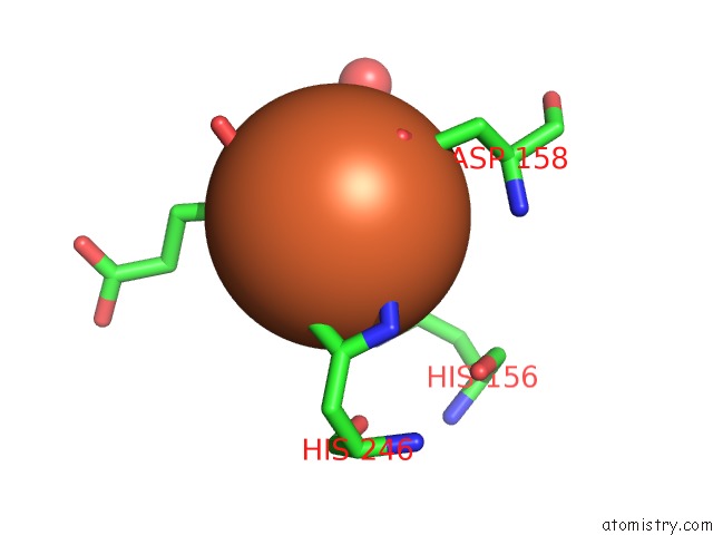
The binding sites of Iron atom in the Crystal Structure of Human Phytanoyl-Coa Dioxygenase PHYHD1 2- Oxoglutarate and Iron Complex
(pdb code 3obz). This binding sites where shown within
5.0 Angstroms radius around Iron atom.
In total only one binding site of Iron was determined in the Crystal Structure of Human Phytanoyl-Coa Dioxygenase PHYHD1 2- Oxoglutarate and Iron Complex, PDB code: 3obz:
In total only one binding site of Iron was determined in the Crystal Structure of Human Phytanoyl-Coa Dioxygenase PHYHD1 2- Oxoglutarate and Iron Complex, PDB code: 3obz:
Iron binding site 1 out of 1 in 3obz
Go back to
Iron binding site 1 out
of 1 in the Crystal Structure of Human Phytanoyl-Coa Dioxygenase PHYHD1 2- Oxoglutarate and Iron Complex
Mono view
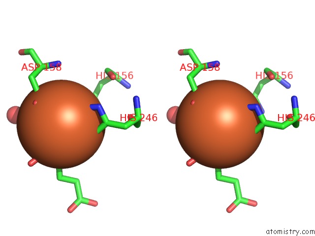
Stereo pair view
Mono view
Stereo pair view
A full contact list of Iron with other atoms in the Fe binding
site number 1 of Crystal Structure of Human Phytanoyl-Coa Dioxygenase PHYHD1 2- Oxoglutarate and Iron Complex within 5.0Å range:
|
Reference:
Z.Zhang,
D.Butler,
M.A.Mcdonough,
K.L.Kavanagh,
J.E.Bray,
S.S.Ng,
F.Von Delft,
C.H.Arrowsmith,
J.Weigelt,
A.Edwards,
M.Sundstrom,
C.J.Schofield,
U.Oppermann.
Crystal Structure of Human PHYHD1, A Non-Heme Fe(II) and 2-Oxoglutarate Dependent Oxygenase Related to Phytanoyl-Coa Hydroxylase To Be Published.
Page generated: Tue Aug 5 05:08:40 2025
Last articles
Na in 1IX0Na in 1IP6
Na in 1IP3
Na in 1IOC
Na in 1IP5
Na in 1IP4
Na in 1IP2
Na in 1IN0
Na in 1IP1
Na in 1IKQ