Iron »
PDB 3om3-3p4q »
3omi »
Iron in PDB 3omi: Catalytic Core Subunits (I and II) of Cytochrome C Oxidase From Rhodobacter Sphaeroides with D132A Mutation
Enzymatic activity of Catalytic Core Subunits (I and II) of Cytochrome C Oxidase From Rhodobacter Sphaeroides with D132A Mutation
All present enzymatic activity of Catalytic Core Subunits (I and II) of Cytochrome C Oxidase From Rhodobacter Sphaeroides with D132A Mutation:
1.9.3.1;
1.9.3.1;
Protein crystallography data
The structure of Catalytic Core Subunits (I and II) of Cytochrome C Oxidase From Rhodobacter Sphaeroides with D132A Mutation, PDB code: 3omi
was solved by
J.Liu,
L.Qin,
S.Ferguson-Miller,
with X-Ray Crystallography technique. A brief refinement statistics is given in the table below:
| Resolution Low / High (Å) | 35.84 / 2.15 |
| Space group | P 21 21 21 |
| Cell size a, b, c (Å), α, β, γ (°) | 125.064, 131.519, 175.674, 90.00, 90.00, 90.00 |
| R / Rfree (%) | 19.2 / 21.5 |
Other elements in 3omi:
The structure of Catalytic Core Subunits (I and II) of Cytochrome C Oxidase From Rhodobacter Sphaeroides with D132A Mutation also contains other interesting chemical elements:
| Magnesium | (Mg) | 2 atoms |
| Cadmium | (Cd) | 4 atoms |
| Calcium | (Ca) | 2 atoms |
| Chlorine | (Cl) | 2 atoms |
| Copper | (Cu) | 6 atoms |
Iron Binding Sites:
The binding sites of Iron atom in the Catalytic Core Subunits (I and II) of Cytochrome C Oxidase From Rhodobacter Sphaeroides with D132A Mutation
(pdb code 3omi). This binding sites where shown within
5.0 Angstroms radius around Iron atom.
In total 4 binding sites of Iron where determined in the Catalytic Core Subunits (I and II) of Cytochrome C Oxidase From Rhodobacter Sphaeroides with D132A Mutation, PDB code: 3omi:
Jump to Iron binding site number: 1; 2; 3; 4;
In total 4 binding sites of Iron where determined in the Catalytic Core Subunits (I and II) of Cytochrome C Oxidase From Rhodobacter Sphaeroides with D132A Mutation, PDB code: 3omi:
Jump to Iron binding site number: 1; 2; 3; 4;
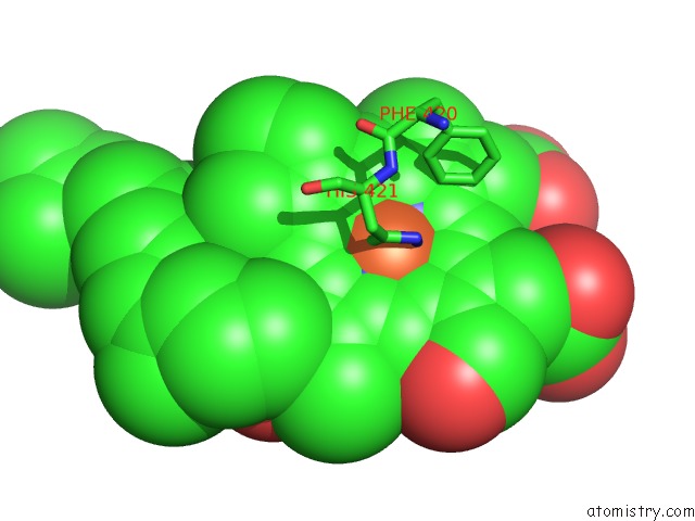
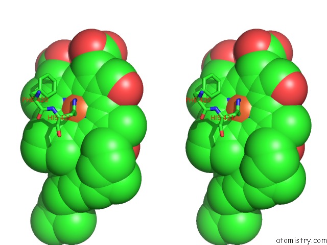
Iron binding site 1 out of 4 in 3omi
Go back to
Iron binding site 1 out
of 4 in the Catalytic Core Subunits (I and II) of Cytochrome C Oxidase From Rhodobacter Sphaeroides with D132A Mutation
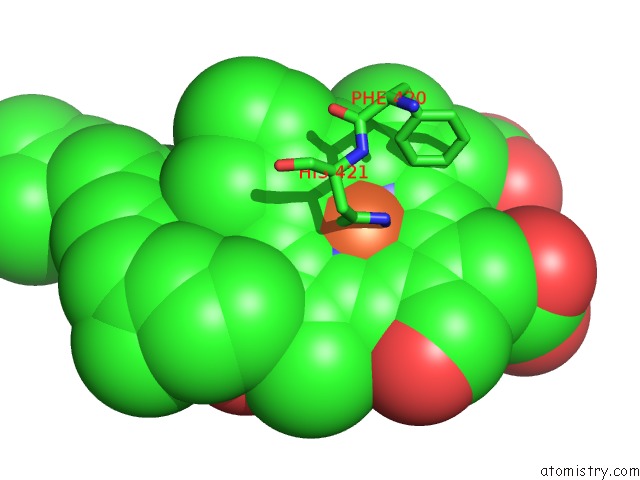
Mono view
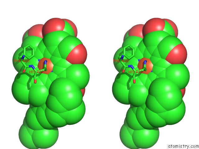
Stereo pair view
Mono view
Stereo pair view
A full contact list of Iron with other atoms in the Fe binding
site number 1 of Catalytic Core Subunits (I and II) of Cytochrome C Oxidase From Rhodobacter Sphaeroides with D132A Mutation within 5.0Å range:
|
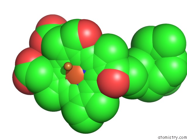
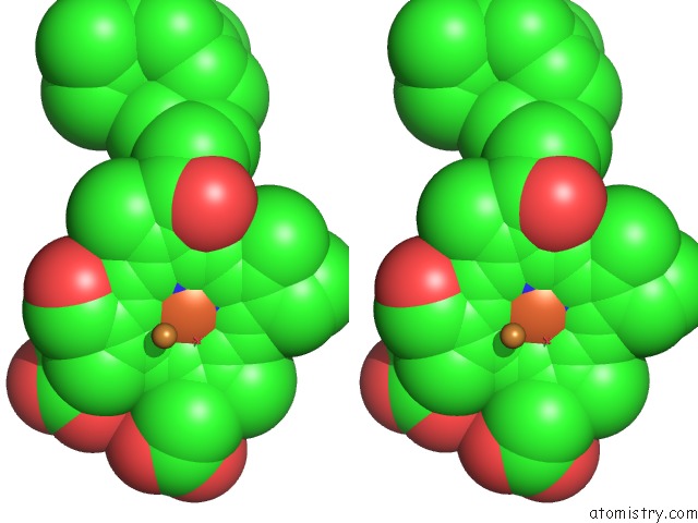
Iron binding site 2 out of 4 in 3omi
Go back to
Iron binding site 2 out
of 4 in the Catalytic Core Subunits (I and II) of Cytochrome C Oxidase From Rhodobacter Sphaeroides with D132A Mutation
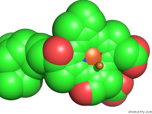
Mono view
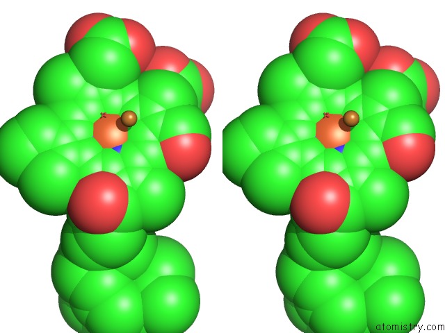
Stereo pair view
Mono view
Stereo pair view
A full contact list of Iron with other atoms in the Fe binding
site number 2 of Catalytic Core Subunits (I and II) of Cytochrome C Oxidase From Rhodobacter Sphaeroides with D132A Mutation within 5.0Å range:
|
Iron binding site 3 out of 4 in 3omi
Go back to
Iron binding site 3 out
of 4 in the Catalytic Core Subunits (I and II) of Cytochrome C Oxidase From Rhodobacter Sphaeroides with D132A Mutation
Mono view
Stereo pair view
Mono view
Stereo pair view
A full contact list of Iron with other atoms in the Fe binding
site number 3 of Catalytic Core Subunits (I and II) of Cytochrome C Oxidase From Rhodobacter Sphaeroides with D132A Mutation within 5.0Å range:
|
Iron binding site 4 out of 4 in 3omi
Go back to
Iron binding site 4 out
of 4 in the Catalytic Core Subunits (I and II) of Cytochrome C Oxidase From Rhodobacter Sphaeroides with D132A Mutation
Mono view
Stereo pair view
Mono view
Stereo pair view
A full contact list of Iron with other atoms in the Fe binding
site number 4 of Catalytic Core Subunits (I and II) of Cytochrome C Oxidase From Rhodobacter Sphaeroides with D132A Mutation within 5.0Å range:
|
Reference:
J.Liu,
L.Qin,
S.Ferguson-Miller.
Crystallographic and Online Spectral Evidence For Role of Conformational Change and Conserved Water in Cytochrome Oxidase Proton Pump. Proc.Natl.Acad.Sci.Usa V. 108 1284 2011.
ISSN: ISSN 0027-8424
PubMed: 21205904
DOI: 10.1073/PNAS.1012846108
Page generated: Tue Aug 5 05:11:39 2025
ISSN: ISSN 0027-8424
PubMed: 21205904
DOI: 10.1073/PNAS.1012846108
Last articles
I in 6B2GI in 6AXY
I in 6AYA
I in 6AY9
I in 6AX6
I in 6AXX
I in 6AXW
I in 6AXT
I in 6AXV
I in 6AXS