Iron »
PDB 3om3-3p4q »
3ovu »
Iron in PDB 3ovu: Crystal Structure of Human Alpha-Haemoglobin Complexed with Ahsp and the First Neat Domain of Isdh From Staphylococcus Aureus
Protein crystallography data
The structure of Crystal Structure of Human Alpha-Haemoglobin Complexed with Ahsp and the First Neat Domain of Isdh From Staphylococcus Aureus, PDB code: 3ovu
was solved by
D.A.Jacques,
K.Krishna Kumar,
T.T.Caradoc-Davies,
D.B.Langley,
J.P.Mackay,
J.M.Guss,
D.A.Gell,
with X-Ray Crystallography technique. A brief refinement statistics is given in the table below:
| Resolution Low / High (Å) | 49.57 / 2.83 |
| Space group | P 21 21 21 |
| Cell size a, b, c (Å), α, β, γ (°) | 68.638, 71.659, 82.058, 90.00, 90.00, 90.00 |
| R / Rfree (%) | 25.2 / 28 |
Iron Binding Sites:
The binding sites of Iron atom in the Crystal Structure of Human Alpha-Haemoglobin Complexed with Ahsp and the First Neat Domain of Isdh From Staphylococcus Aureus
(pdb code 3ovu). This binding sites where shown within
5.0 Angstroms radius around Iron atom.
In total only one binding site of Iron was determined in the Crystal Structure of Human Alpha-Haemoglobin Complexed with Ahsp and the First Neat Domain of Isdh From Staphylococcus Aureus, PDB code: 3ovu:
In total only one binding site of Iron was determined in the Crystal Structure of Human Alpha-Haemoglobin Complexed with Ahsp and the First Neat Domain of Isdh From Staphylococcus Aureus, PDB code: 3ovu:
Iron binding site 1 out of 1 in 3ovu
Go back to
Iron binding site 1 out
of 1 in the Crystal Structure of Human Alpha-Haemoglobin Complexed with Ahsp and the First Neat Domain of Isdh From Staphylococcus Aureus
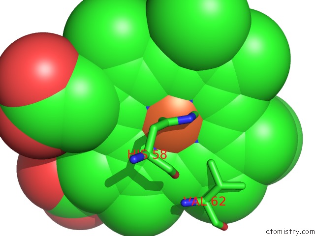
Mono view
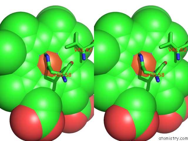
Stereo pair view
Mono view
Stereo pair view
A full contact list of Iron with other atoms in the Fe binding
site number 1 of Crystal Structure of Human Alpha-Haemoglobin Complexed with Ahsp and the First Neat Domain of Isdh From Staphylococcus Aureus within 5.0Å range:
|
Reference:
K.Krishna Kumar,
D.A.Jacques,
T.T.Caradoc-Davies,
T.Spirig,
D.B.Langley,
J.P.Mackay,
J.M.Guss,
R.T.Clubb,
D.A.Gell.
A New Haem Pocket Structure in Alpha-Haemoglobin To Be Published.
Page generated: Sun Aug 4 17:18:27 2024
Last articles
Fe in 2YXOFe in 2YRS
Fe in 2YXC
Fe in 2YNM
Fe in 2YVJ
Fe in 2YP1
Fe in 2YU2
Fe in 2YU1
Fe in 2YQB
Fe in 2YOO