Iron »
PDB 3om3-3p4q »
3p3z »
Iron in PDB 3p3z: Crystal Structure of the Cytochrome P450 Monooxygenase Aurh From Streptomyces Thioluteus in Complex with Ancymidol
Protein crystallography data
The structure of Crystal Structure of the Cytochrome P450 Monooxygenase Aurh From Streptomyces Thioluteus in Complex with Ancymidol, PDB code: 3p3z
was solved by
G.Zocher,
M.E.A.Richter,
U.Mueller,
C.Hertweck,
with X-Ray Crystallography technique. A brief refinement statistics is given in the table below:
| Resolution Low / High (Å) | 20.00 / 2.30 |
| Space group | H 3 |
| Cell size a, b, c (Å), α, β, γ (°) | 129.370, 129.370, 71.070, 90.00, 90.00, 120.00 |
| R / Rfree (%) | 18.3 / 23.1 |
Other elements in 3p3z:
The structure of Crystal Structure of the Cytochrome P450 Monooxygenase Aurh From Streptomyces Thioluteus in Complex with Ancymidol also contains other interesting chemical elements:
| Chlorine | (Cl) | 3 atoms |
Iron Binding Sites:
The binding sites of Iron atom in the Crystal Structure of the Cytochrome P450 Monooxygenase Aurh From Streptomyces Thioluteus in Complex with Ancymidol
(pdb code 3p3z). This binding sites where shown within
5.0 Angstroms radius around Iron atom.
In total only one binding site of Iron was determined in the Crystal Structure of the Cytochrome P450 Monooxygenase Aurh From Streptomyces Thioluteus in Complex with Ancymidol, PDB code: 3p3z:
In total only one binding site of Iron was determined in the Crystal Structure of the Cytochrome P450 Monooxygenase Aurh From Streptomyces Thioluteus in Complex with Ancymidol, PDB code: 3p3z:
Iron binding site 1 out of 1 in 3p3z
Go back to
Iron binding site 1 out
of 1 in the Crystal Structure of the Cytochrome P450 Monooxygenase Aurh From Streptomyces Thioluteus in Complex with Ancymidol
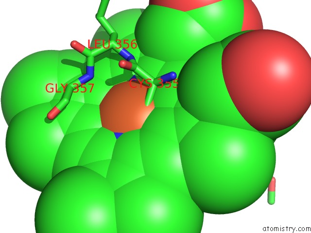
Mono view
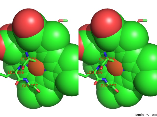
Stereo pair view
Mono view
Stereo pair view
A full contact list of Iron with other atoms in the Fe binding
site number 1 of Crystal Structure of the Cytochrome P450 Monooxygenase Aurh From Streptomyces Thioluteus in Complex with Ancymidol within 5.0Å range:
|
Reference:
G.Zocher,
M.E.Richter,
U.Mueller,
C.Hertweck.
Structural Fine-Tuning of A Multifunctional Cytochrome P450 Monooxygenase. J.Am.Chem.Soc. V. 133 2292 2011.
ISSN: ISSN 0002-7863
PubMed: 21280577
DOI: 10.1021/JA110146Z
Page generated: Sun Aug 4 17:24:07 2024
ISSN: ISSN 0002-7863
PubMed: 21280577
DOI: 10.1021/JA110146Z
Last articles
Fe in 2YXOFe in 2YRS
Fe in 2YXC
Fe in 2YNM
Fe in 2YVJ
Fe in 2YP1
Fe in 2YU2
Fe in 2YU1
Fe in 2YQB
Fe in 2YOO