Iron »
PDB 3p4r-3pcn »
3p5q »
Iron in PDB 3p5q: Ferric R-State Human Aquomethemoglobin
Protein crystallography data
The structure of Ferric R-State Human Aquomethemoglobin, PDB code: 3p5q
was solved by
J.Yi,
L.M.Thomas,
G.B.Richter-Addo,
with X-Ray Crystallography technique. A brief refinement statistics is given in the table below:
| Resolution Low / High (Å) | 18.14 / 2.00 |
| Space group | P 41 21 2 |
| Cell size a, b, c (Å), α, β, γ (°) | 53.546, 53.546, 192.760, 90.00, 90.00, 90.00 |
| R / Rfree (%) | 22.1 / 26.7 |
Iron Binding Sites:
The binding sites of Iron atom in the Ferric R-State Human Aquomethemoglobin
(pdb code 3p5q). This binding sites where shown within
5.0 Angstroms radius around Iron atom.
In total 2 binding sites of Iron where determined in the Ferric R-State Human Aquomethemoglobin, PDB code: 3p5q:
Jump to Iron binding site number: 1; 2;
In total 2 binding sites of Iron where determined in the Ferric R-State Human Aquomethemoglobin, PDB code: 3p5q:
Jump to Iron binding site number: 1; 2;
Iron binding site 1 out of 2 in 3p5q
Go back to
Iron binding site 1 out
of 2 in the Ferric R-State Human Aquomethemoglobin
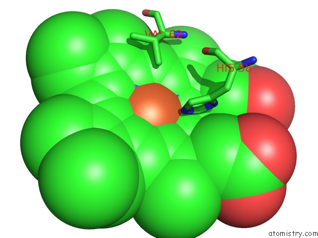
Mono view
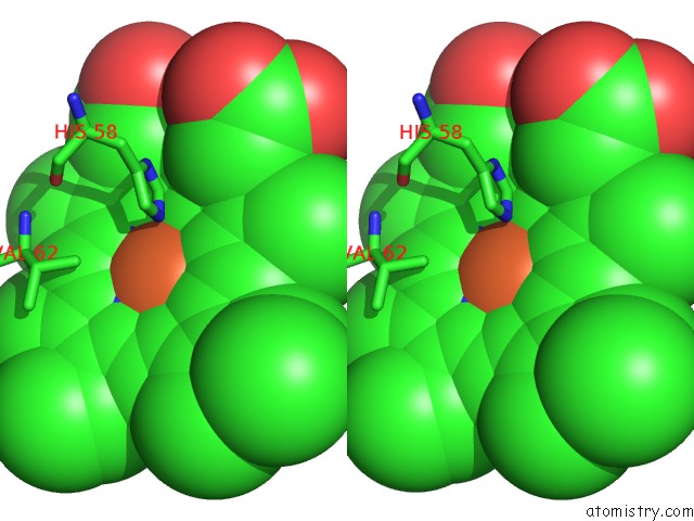
Stereo pair view
Mono view
Stereo pair view
A full contact list of Iron with other atoms in the Fe binding
site number 1 of Ferric R-State Human Aquomethemoglobin within 5.0Å range:
|
Iron binding site 2 out of 2 in 3p5q
Go back to
Iron binding site 2 out
of 2 in the Ferric R-State Human Aquomethemoglobin
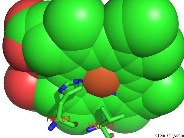
Mono view
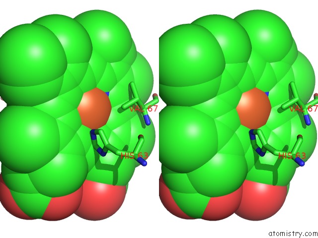
Stereo pair view
Mono view
Stereo pair view
A full contact list of Iron with other atoms in the Fe binding
site number 2 of Ferric R-State Human Aquomethemoglobin within 5.0Å range:
|
Reference:
J.Yi,
L.M.Thomas,
G.B.Richter-Addo.
Structure of Human R-State Aquomethemoglobin at 2.0 A Resolution Acta Crystallogr.,Sect.F V. 67 647 2011.
ISSN: ESSN 1744-3091
PubMed: 21636902
DOI: 10.1107/S1744309111012528
Page generated: Tue Aug 5 05:28:27 2025
ISSN: ESSN 1744-3091
PubMed: 21636902
DOI: 10.1107/S1744309111012528
Last articles
Yb in 1YTTYb in 2O6N
Yb in 2K62
Yb in 2BOP
Yb in 1PK0
Yb in 1S26
Yb in 1SQM
Yb in 1GGY
Yb in 1NCH
Yb in 1LVC