Iron »
PDB 3p4r-3pcn »
3pah »
Iron in PDB 3pah: Human Phenylalanine Hydroxylase Catalytic Domain Dimer with Bound Adrenaline Inhibitor
Enzymatic activity of Human Phenylalanine Hydroxylase Catalytic Domain Dimer with Bound Adrenaline Inhibitor
All present enzymatic activity of Human Phenylalanine Hydroxylase Catalytic Domain Dimer with Bound Adrenaline Inhibitor:
1.14.16.1;
1.14.16.1;
Protein crystallography data
The structure of Human Phenylalanine Hydroxylase Catalytic Domain Dimer with Bound Adrenaline Inhibitor, PDB code: 3pah
was solved by
H.Erlandsen,
T.Flatmark,
R.C.Stevens,
with X-Ray Crystallography technique. A brief refinement statistics is given in the table below:
| Resolution Low / High (Å) | 20.00 / 2.00 |
| Space group | C 2 2 21 |
| Cell size a, b, c (Å), α, β, γ (°) | 66.700, 108.650, 125.470, 90.00, 90.00, 90.00 |
| R / Rfree (%) | 17.5 / 20.4 |
Iron Binding Sites:
The binding sites of Iron atom in the Human Phenylalanine Hydroxylase Catalytic Domain Dimer with Bound Adrenaline Inhibitor
(pdb code 3pah). This binding sites where shown within
5.0 Angstroms radius around Iron atom.
In total only one binding site of Iron was determined in the Human Phenylalanine Hydroxylase Catalytic Domain Dimer with Bound Adrenaline Inhibitor, PDB code: 3pah:
In total only one binding site of Iron was determined in the Human Phenylalanine Hydroxylase Catalytic Domain Dimer with Bound Adrenaline Inhibitor, PDB code: 3pah:
Iron binding site 1 out of 1 in 3pah
Go back to
Iron binding site 1 out
of 1 in the Human Phenylalanine Hydroxylase Catalytic Domain Dimer with Bound Adrenaline Inhibitor
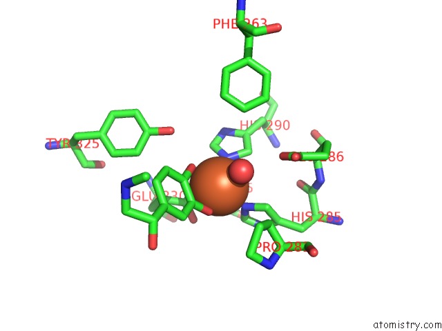
Mono view
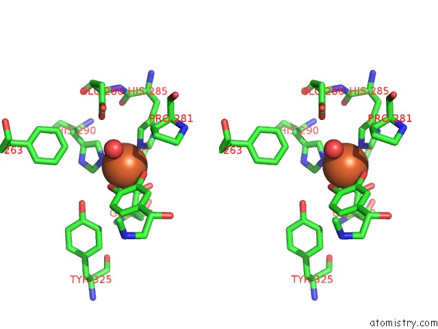
Stereo pair view
Mono view
Stereo pair view
A full contact list of Iron with other atoms in the Fe binding
site number 1 of Human Phenylalanine Hydroxylase Catalytic Domain Dimer with Bound Adrenaline Inhibitor within 5.0Å range:
|
Reference:
H.Erlandsen,
T.Flatmark,
R.C.Stevens,
E.Hough.
Crystallographic Analysis of the Human Phenylalanine Hydroxylase Catalytic Domain with Bound Catechol Inhibitors at 2.0 A Resolution. Biochemistry V. 37 15638 1998.
ISSN: ISSN 0006-2960
PubMed: 9843368
DOI: 10.1021/BI9815290
Page generated: Tue Aug 5 05:35:12 2025
ISSN: ISSN 0006-2960
PubMed: 9843368
DOI: 10.1021/BI9815290
Last articles
Yb in 1YTTYb in 2O6N
Yb in 2K62
Yb in 2BOP
Yb in 1PK0
Yb in 1S26
Yb in 1SQM
Yb in 1GGY
Yb in 1NCH
Yb in 1LVC