Iron »
PDB 3p4r-3pcn »
3pcf »
Iron in PDB 3pcf: Structure of Protocatechuate 3,4-Dioxygenase Complexed with 3-Fluro-4- Hydroxybenzoate
Enzymatic activity of Structure of Protocatechuate 3,4-Dioxygenase Complexed with 3-Fluro-4- Hydroxybenzoate
All present enzymatic activity of Structure of Protocatechuate 3,4-Dioxygenase Complexed with 3-Fluro-4- Hydroxybenzoate:
1.13.11.3;
1.13.11.3;
Protein crystallography data
The structure of Structure of Protocatechuate 3,4-Dioxygenase Complexed with 3-Fluro-4- Hydroxybenzoate, PDB code: 3pcf
was solved by
A.M.Orville,
N.Elango,
J.D.Lipscomb,
D.H.Ohlendorf,
with X-Ray Crystallography technique. A brief refinement statistics is given in the table below:
| Resolution Low / High (Å) | 6.00 / 2.15 |
| Space group | I 1 2 1 |
| Cell size a, b, c (Å), α, β, γ (°) | 196.030, 127.220, 133.700, 90.00, 97.70, 90.00 |
| R / Rfree (%) | n/a / n/a |
Other elements in 3pcf:
The structure of Structure of Protocatechuate 3,4-Dioxygenase Complexed with 3-Fluro-4- Hydroxybenzoate also contains other interesting chemical elements:
| Fluorine | (F) | 12 atoms |
Iron Binding Sites:
The binding sites of Iron atom in the Structure of Protocatechuate 3,4-Dioxygenase Complexed with 3-Fluro-4- Hydroxybenzoate
(pdb code 3pcf). This binding sites where shown within
5.0 Angstroms radius around Iron atom.
In total 6 binding sites of Iron where determined in the Structure of Protocatechuate 3,4-Dioxygenase Complexed with 3-Fluro-4- Hydroxybenzoate, PDB code: 3pcf:
Jump to Iron binding site number: 1; 2; 3; 4; 5; 6;
In total 6 binding sites of Iron where determined in the Structure of Protocatechuate 3,4-Dioxygenase Complexed with 3-Fluro-4- Hydroxybenzoate, PDB code: 3pcf:
Jump to Iron binding site number: 1; 2; 3; 4; 5; 6;
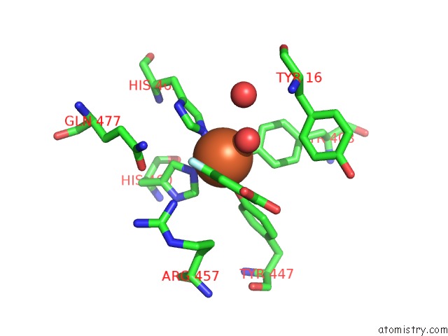
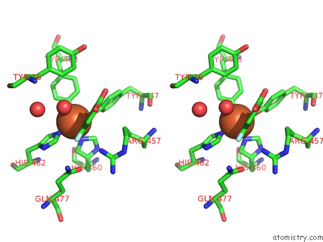
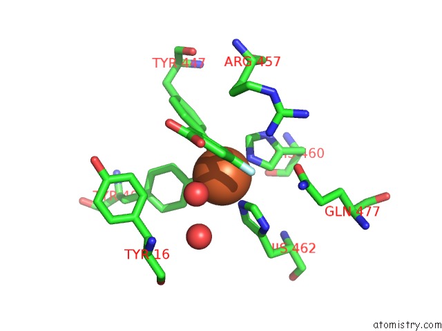
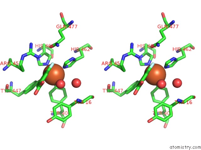
Iron binding site 1 out of 6 in 3pcf
Go back to
Iron binding site 1 out
of 6 in the Structure of Protocatechuate 3,4-Dioxygenase Complexed with 3-Fluro-4- Hydroxybenzoate
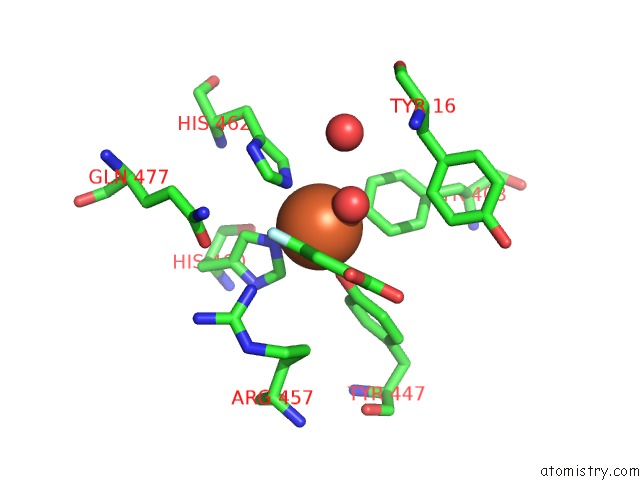
Mono view
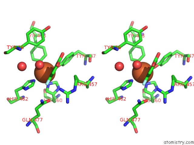
Stereo pair view
Mono view
Stereo pair view
A full contact list of Iron with other atoms in the Fe binding
site number 1 of Structure of Protocatechuate 3,4-Dioxygenase Complexed with 3-Fluro-4- Hydroxybenzoate within 5.0Å range:
|
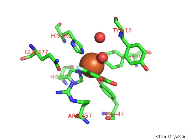
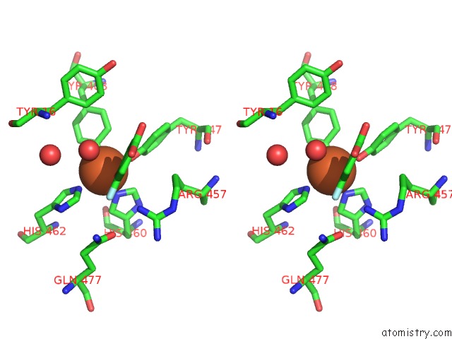
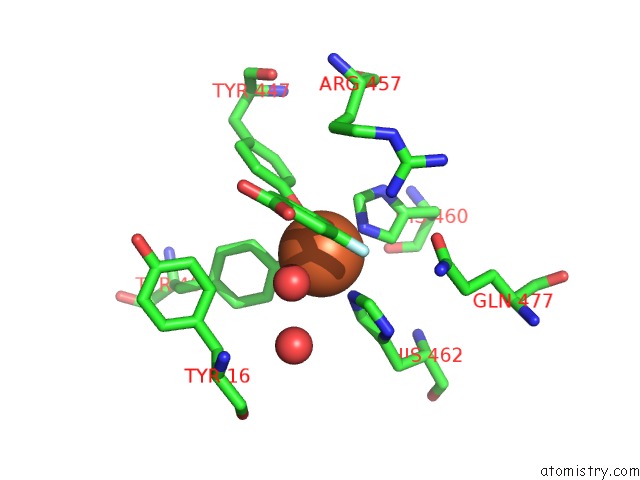
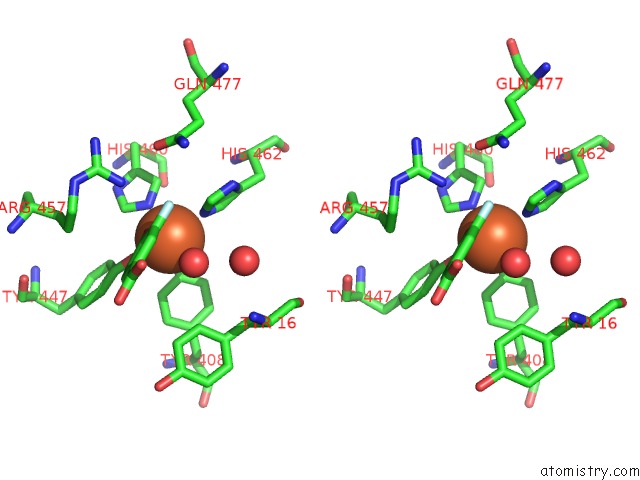
Iron binding site 2 out of 6 in 3pcf
Go back to
Iron binding site 2 out
of 6 in the Structure of Protocatechuate 3,4-Dioxygenase Complexed with 3-Fluro-4- Hydroxybenzoate
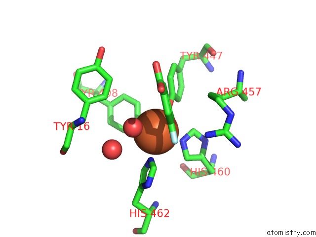
Mono view
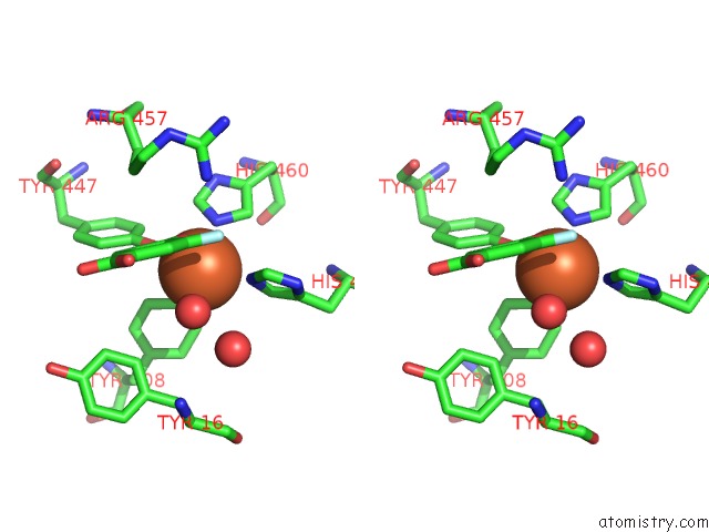
Stereo pair view
Mono view
Stereo pair view
A full contact list of Iron with other atoms in the Fe binding
site number 2 of Structure of Protocatechuate 3,4-Dioxygenase Complexed with 3-Fluro-4- Hydroxybenzoate within 5.0Å range:
|
Iron binding site 3 out of 6 in 3pcf
Go back to
Iron binding site 3 out
of 6 in the Structure of Protocatechuate 3,4-Dioxygenase Complexed with 3-Fluro-4- Hydroxybenzoate
Mono view
Stereo pair view
Mono view
Stereo pair view
A full contact list of Iron with other atoms in the Fe binding
site number 3 of Structure of Protocatechuate 3,4-Dioxygenase Complexed with 3-Fluro-4- Hydroxybenzoate within 5.0Å range:
|
Iron binding site 4 out of 6 in 3pcf
Go back to
Iron binding site 4 out
of 6 in the Structure of Protocatechuate 3,4-Dioxygenase Complexed with 3-Fluro-4- Hydroxybenzoate
Mono view
Stereo pair view
Mono view
Stereo pair view
A full contact list of Iron with other atoms in the Fe binding
site number 4 of Structure of Protocatechuate 3,4-Dioxygenase Complexed with 3-Fluro-4- Hydroxybenzoate within 5.0Å range:
|
Iron binding site 5 out of 6 in 3pcf
Go back to
Iron binding site 5 out
of 6 in the Structure of Protocatechuate 3,4-Dioxygenase Complexed with 3-Fluro-4- Hydroxybenzoate
Mono view
Stereo pair view
Mono view
Stereo pair view
A full contact list of Iron with other atoms in the Fe binding
site number 5 of Structure of Protocatechuate 3,4-Dioxygenase Complexed with 3-Fluro-4- Hydroxybenzoate within 5.0Å range:
|
Iron binding site 6 out of 6 in 3pcf
Go back to
Iron binding site 6 out
of 6 in the Structure of Protocatechuate 3,4-Dioxygenase Complexed with 3-Fluro-4- Hydroxybenzoate
Mono view
Stereo pair view
Mono view
Stereo pair view
A full contact list of Iron with other atoms in the Fe binding
site number 6 of Structure of Protocatechuate 3,4-Dioxygenase Complexed with 3-Fluro-4- Hydroxybenzoate within 5.0Å range:
|
Reference:
A.M.Orville,
N.Elango,
J.D.Lipscomb,
D.H.Ohlendorf.
Structures of Competitive Inhibitor Complexes of Protocatechuate 3,4-Dioxygenase: Multiple Exogenous Ligand Binding Orientations Within the Active Site. Biochemistry V. 36 10039 1997.
ISSN: ISSN 0006-2960
PubMed: 9254599
DOI: 10.1021/BI970468N
Page generated: Tue Aug 5 05:37:56 2025
ISSN: ISSN 0006-2960
PubMed: 9254599
DOI: 10.1021/BI970468N
Last articles
Yb in 1NCGYb in 1HS6
Yb in 1H19
Yb in 1C5K
Yb in 1GW6
Yb in 1CNT
Y in 8PUN
Y in 6PRF
Y in 8U7C
Y in 6NDG