Atomistry » Iron » PDB 3pur-3qjs » 3pwf
Atomistry »
  Iron »
    PDB 3pur-3qjs »
      3pwf »

Iron in PDB 3pwf: High Resolution Structure of the Fully Reduced Form of Rubrerythrin From P. Furiosus

Protein crystallography data

The structure of High Resolution Structure of the Fully Reduced Form of Rubrerythrin From P. Furiosus, PDB code: 3pwf was solved by B.D.Dillard, J.M.Demick, M.W.Adams, W.N.Lanzilotta, with X-Ray Crystallography technique. A brief refinement statistics is given in the table below:

Resolution Low / High (Å) 33.63 / 1.64
Space group P 42 21 2
Cell size a, b, c (Å), α, β, γ (°) 104.870, 104.870, 79.840, 90.00, 90.00, 90.00
R / Rfree (%) 16.6 / 18.8

Iron Binding Sites:

The binding sites of Iron atom in the High Resolution Structure of the Fully Reduced Form of Rubrerythrin From P. Furiosus (pdb code 3pwf). This binding sites where shown within 5.0 Angstroms radius around Iron atom.
In total 6 binding sites of Iron where determined in the High Resolution Structure of the Fully Reduced Form of Rubrerythrin From P. Furiosus, PDB code: 3pwf:
Jump to Iron binding site number: 1; 2; 3; 4; 5; 6;

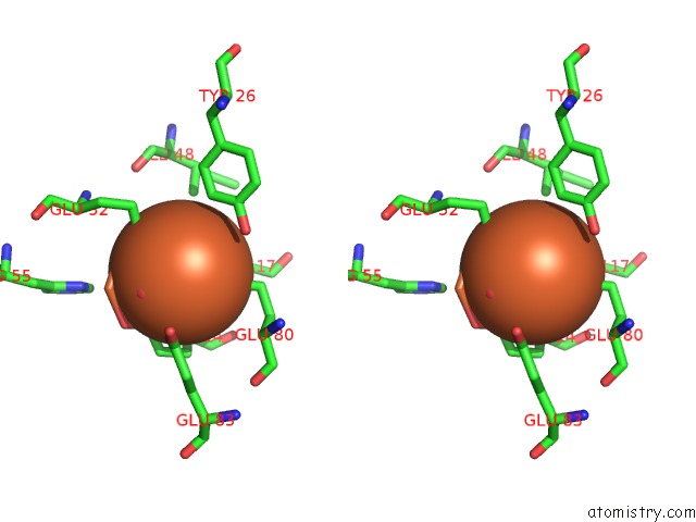
Iron binding site 1 out of 6 in 3pwf

Go back to Iron Binding Sites List in 3pwf
Iron binding site 1 out of 6 in the High Resolution Structure of the Fully Reduced Form of Rubrerythrin From P. Furiosus


Mono view


Stereo pair view

A full contact list of Iron with other atoms in the Fe binding site number 1 of High Resolution Structure of the Fully Reduced Form of Rubrerythrin From P. Furiosus within 5.0Å range:
probe atom residue distance (Å) B Occ
A:Fe200

b:12.8
occ:1.00
SG A:CYS160 2.3 17.1 1.0
SG A:CYS145 2.3 12.3 1.0
SG A:CYS157 2.3 14.2 1.0
SG A:CYS142 2.4 14.1 1.0
CB A:CYS142 3.2 12.4 1.0
CB A:CYS157 3.3 11.3 1.0
CB A:CYS160 3.5 17.2 1.0
CB A:CYS145 3.5 12.0 1.0
N A:CYS160 3.8 14.6 1.0
N A:CYS145 3.9 11.0 1.0
CA A:CYS145 4.2 11.7 1.0
CA A:CYS160 4.2 16.3 1.0
CB A:TYR147 4.3 10.7 1.0
CB A:ILE144 4.4 13.7 1.0
CB A:ALA162 4.6 16.4 1.0
CA A:CYS157 4.7 13.3 1.0
CB A:VAL159 4.7 12.3 1.0
CA A:CYS142 4.7 11.4 1.0
C A:ILE144 4.8 14.0 1.0
C A:CYS160 4.8 16.4 1.0
C A:CYS145 4.8 12.3 1.0
N A:TYR147 4.8 11.9 1.0
C A:VAL159 4.9 13.4 1.0
N A:GLY161 4.9 15.8 1.0
N A:GLY146 4.9 12.0 1.0
N A:ALA162 4.9 15.8 1.0
CG2 A:ILE144 4.9 13.7 1.0
CA A:ILE144 5.0 13.3 1.0
N A:ILE144 5.0 13.0 1.0

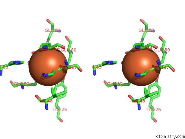
Iron binding site 2 out of 6 in 3pwf

Go back to Iron Binding Sites List in 3pwf
Iron binding site 2 out of 6 in the High Resolution Structure of the Fully Reduced Form of Rubrerythrin From P. Furiosus


Mono view


Stereo pair view

A full contact list of Iron with other atoms in the Fe binding site number 2 of High Resolution Structure of the Fully Reduced Form of Rubrerythrin From P. Furiosus within 5.0Å range:
probe atom residue distance (Å) B Occ
A:Fe201

b:11.7
occ:1.00
OE2 B:GLU52 2.0 12.3 1.0
OE2 A:GLU114 2.0 12.0 1.0
OE1 A:GLU80 2.1 13.4 1.0
OE2 A:GLU80 2.1 11.4 1.0
ND1 A:HIS117 2.1 9.6 1.0
O A:HOH288 2.2 25.0 1.0
CD A:GLU80 2.5 12.1 1.0
O A:HOH176 2.9 21.2 1.0
CD B:GLU52 2.9 12.6 1.0
CE1 A:HIS117 3.0 10.5 1.0
CD A:GLU114 3.0 10.7 1.0
OE1 B:GLU52 3.2 12.2 1.0
CG A:HIS117 3.3 10.0 1.0
CB A:HIS117 3.7 10.7 1.0
FE B:FE2202 3.8 10.1 1.0
OE1 A:GLU114 3.8 11.8 1.0
CG A:GLU80 4.0 12.5 1.0
CA A:GLU114 4.1 9.0 1.0
CG A:GLU114 4.1 9.6 1.0
CB A:GLU114 4.2 8.5 1.0
NE2 A:HIS117 4.3 9.8 1.0
OE2 A:GLU83 4.3 20.4 1.0
CG B:GLU52 4.3 9.9 1.0
CG2 B:ILE48 4.4 11.0 1.0
CD2 A:HIS117 4.4 10.1 1.0
CE1 B:TYR26 4.5 9.8 1.0
OH B:TYR26 4.6 13.4 1.0
N A:GLU114 4.8 12.1 1.0
CB A:GLU80 4.9 9.7 1.0
CE1 B:HIS55 5.0 12.5 1.0

Iron binding site 3 out of 6 in 3pwf

Go back to Iron Binding Sites List in 3pwf
Iron binding site 3 out of 6 in the High Resolution Structure of the Fully Reduced Form of Rubrerythrin From P. Furiosus


Mono view


Stereo pair view

A full contact list of Iron with other atoms in the Fe binding site number 3 of High Resolution Structure of the Fully Reduced Form of Rubrerythrin From P. Furiosus within 5.0Å range:
probe atom residue distance (Å) B Occ
A:Fe202

b:10.5
occ:1.00
OE2 A:GLU52 2.0 13.0 1.0
OE1 B:GLU114 2.0 12.0 1.0
OE2 A:GLU19 2.1 10.8 1.0
ND1 A:HIS55 2.2 9.8 1.0
OE1 A:GLU19 2.2 12.8 1.0
O B:HOH172 2.3 21.9 1.0
CD A:GLU19 2.5 11.9 1.0
CD B:GLU114 2.9 11.5 1.0
CE1 A:HIS55 3.0 10.8 1.0
OE2 B:GLU114 3.0 12.7 1.0
CD A:GLU52 3.0 12.6 1.0
CG A:HIS55 3.3 8.8 1.0
OE1 A:GLU52 3.7 12.2 1.0
CB A:HIS55 3.7 8.6 1.0
FE B:FE2201 3.8 12.3 1.0
O B:HOH290 3.9 28.6 1.0
CG A:GLU19 4.1 11.0 1.0
CA A:GLU52 4.2 10.6 1.0
NE2 A:HIS55 4.2 10.9 1.0
O B:HOH191 4.2 42.0 1.0
CG A:GLU52 4.3 11.7 1.0
CG B:GLU114 4.3 12.5 1.0
OE1 B:GLU83 4.3 23.2 1.0
CB A:GLU52 4.3 10.1 1.0
CD2 A:HIS55 4.4 12.2 1.0
CB B:ALA110 4.5 10.7 1.0
OH B:TYR88 4.7 10.0 1.0
CE2 B:TYR88 4.8 10.2 1.0
CB A:GLU19 4.9 10.4 1.0
CE1 B:HIS117 4.9 12.1 1.0
O B:ALA110 5.0 11.7 1.0
N A:GLU52 5.0 10.5 1.0

Iron binding site 4 out of 6 in 3pwf

Go back to Iron Binding Sites List in 3pwf
Iron binding site 4 out of 6 in the High Resolution Structure of the Fully Reduced Form of Rubrerythrin From P. Furiosus


Mono view


Stereo pair view

A full contact list of Iron with other atoms in the Fe binding site number 4 of High Resolution Structure of the Fully Reduced Form of Rubrerythrin From P. Furiosus within 5.0Å range:
probe atom residue distance (Å) B Occ
B:Fe200

b:12.9
occ:1.00
SG B:CYS145 2.3 13.0 1.0
SG B:CYS160 2.3 14.8 1.0
SG B:CYS157 2.4 14.9 1.0
SG B:CYS142 2.4 14.3 1.0
CB B:CYS142 3.2 12.3 1.0
CB B:CYS157 3.4 12.1 1.0
CB B:CYS160 3.4 15.2 1.0
CB B:CYS145 3.6 13.0 1.0
N B:CYS160 3.8 15.1 1.0
N B:CYS145 3.8 13.0 1.0
CA B:CYS145 4.2 14.0 1.0
CA B:CYS160 4.2 12.5 1.0
CB B:TYR147 4.2 12.8 1.0
CB B:ILE144 4.3 15.8 1.0
CB B:ALA162 4.6 13.1 1.0
CA B:CYS142 4.7 10.3 1.0
CB B:VAL159 4.7 12.4 1.0
CA B:CYS157 4.7 13.5 1.0
C B:ILE144 4.7 16.2 1.0
N B:TYR147 4.8 13.8 1.0
C B:VAL159 4.8 13.8 1.0
C B:CYS160 4.9 14.1 1.0
N B:GLY161 4.9 14.1 1.0
N B:GLY146 4.9 13.0 1.0
C B:CYS145 4.9 15.3 1.0
N B:ALA162 4.9 13.6 1.0
CG2 B:ILE144 4.9 19.6 1.0
CA B:ILE144 4.9 15.6 1.0

Iron binding site 5 out of 6 in 3pwf

Go back to Iron Binding Sites List in 3pwf
Iron binding site 5 out of 6 in the High Resolution Structure of the Fully Reduced Form of Rubrerythrin From P. Furiosus


Mono view


Stereo pair view

A full contact list of Iron with other atoms in the Fe binding site number 5 of High Resolution Structure of the Fully Reduced Form of Rubrerythrin From P. Furiosus within 5.0Å range:
probe atom residue distance (Å) B Occ
B:Fe201

b:12.3
occ:1.00
OE2 B:GLU114 2.0 12.7 1.0
OE1 A:GLU52 2.0 12.2 1.0
OE2 B:GLU80 2.1 15.3 1.0
OE1 B:GLU80 2.1 12.4 1.0
ND1 B:HIS117 2.2 10.2 1.0
O B:HOH290 2.3 28.6 1.0
CD B:GLU80 2.5 16.6 1.0
O B:HOH172 2.9 21.9 1.0
CD A:GLU52 2.9 12.6 1.0
CD B:GLU114 3.0 11.5 1.0
CE1 B:HIS117 3.1 12.1 1.0
OE2 A:GLU52 3.2 13.0 1.0
CG B:HIS117 3.3 10.2 1.0
CB B:HIS117 3.7 13.1 1.0
FE A:FE2202 3.8 10.5 1.0
OE1 B:GLU114 3.8 12.0 1.0
CG B:GLU80 4.0 11.6 1.0
CA B:GLU114 4.1 10.7 1.0
CG B:GLU114 4.1 12.5 1.0
CB B:GLU114 4.2 9.4 1.0
OE1 B:GLU83 4.3 23.2 1.0
NE2 B:HIS117 4.3 12.1 1.0
CG A:GLU52 4.3 11.7 1.0
CG2 A:ILE48 4.3 12.3 1.0
CD2 B:HIS117 4.4 11.9 1.0
OH A:TYR26 4.5 14.9 1.0
CE1 A:TYR26 4.6 10.6 1.0
N B:GLU114 4.8 10.1 1.0
CB B:GLU80 4.9 11.1 1.0
CE1 A:HIS55 4.9 10.8 1.0

Iron binding site 6 out of 6 in 3pwf

Go back to Iron Binding Sites List in 3pwf
Iron binding site 6 out of 6 in the High Resolution Structure of the Fully Reduced Form of Rubrerythrin From P. Furiosus


Mono view


Stereo pair view

A full contact list of Iron with other atoms in the Fe binding site number 6 of High Resolution Structure of the Fully Reduced Form of Rubrerythrin From P. Furiosus within 5.0Å range:
probe atom residue distance (Å) B Occ
B:Fe202

b:10.1
occ:1.00
OE1 B:GLU52 2.0 12.2 1.0
OE1 A:GLU114 2.0 11.8 1.0
OE1 B:GLU19 2.1 10.8 1.0
ND1 B:HIS55 2.2 10.7 1.0
OE2 B:GLU19 2.2 10.5 1.0
O A:HOH176 2.2 21.2 1.0
CD B:GLU19 2.5 8.6 1.0
CD A:GLU114 2.9 10.7 1.0
OE2 A:GLU114 3.0 12.0 1.0
CD B:GLU52 3.0 12.6 1.0
CE1 B:HIS55 3.1 12.5 1.0
CG B:HIS55 3.3 9.8 1.0
OE2 B:GLU52 3.7 12.3 1.0
CB B:HIS55 3.7 10.2 1.0
FE A:FE2201 3.8 11.7 1.0
O A:HOH288 3.9 25.0 1.0
CG B:GLU19 4.1 10.7 1.0
CA B:GLU52 4.1 10.4 1.0
OE2 A:GLU83 4.2 20.4 1.0
CG B:GLU52 4.2 9.9 1.0
O A:HOH278 4.2 35.1 1.0
NE2 B:HIS55 4.3 11.6 1.0
CG A:GLU114 4.3 9.6 1.0
CB B:GLU52 4.3 9.7 1.0
CD2 B:HIS55 4.4 10.0 1.0
CB A:ALA110 4.6 10.2 1.0
OH A:TYR88 4.7 10.4 1.0
CE2 A:TYR88 4.8 10.4 1.0
CE1 A:HIS117 4.9 10.5 1.0
CB B:GLU19 4.9 8.7 1.0
O B:ALA51 4.9 10.1 1.0
N B:GLU52 4.9 10.4 1.0
O A:ALA110 5.0 11.4 1.0
ND1 A:HIS117 5.0 9.6 1.0

Reference:

B.D.Dillard, J.M.Demick, M.W.Adams, W.N.Lanzilotta. A Cryo-Crystallographic Time Course For Peroxide Reduction By Rubrerythrin From Pyrococcus Furiosus. J.Biol.Inorg.Chem. V. 16 949 2011.
ISSN: ISSN 0949-8257
PubMed: 21647777
DOI: 10.1007/S00775-011-0795-6
Page generated: Tue Aug 5 05:56:48 2025

Last articles

Mn in 2GU6
Mn in 2GU5
Mn in 2GU4
Mn in 2FFL
Mn in 2GTX
Mn in 2GNM
Mn in 2GND
Mn in 2GMV
Mn in 2GLF
Mn in 2GLK
© Copyright 2008-2020 by atomistry.com
Home   |    Site Map   |    Copyright   |    Contact us   |    Privacy