Iron »
PDB 3r1a-3rmk »
3r1j »
Iron in PDB 3r1j: Crystal Structure of Alpha-Ketoglutarate-Dependent Taurine Dioxygenase From Mycobacterium Avium, Native Form
Enzymatic activity of Crystal Structure of Alpha-Ketoglutarate-Dependent Taurine Dioxygenase From Mycobacterium Avium, Native Form
All present enzymatic activity of Crystal Structure of Alpha-Ketoglutarate-Dependent Taurine Dioxygenase From Mycobacterium Avium, Native Form:
1.14.11.17;
1.14.11.17;
Protein crystallography data
The structure of Crystal Structure of Alpha-Ketoglutarate-Dependent Taurine Dioxygenase From Mycobacterium Avium, Native Form, PDB code: 3r1j
was solved by
Seattle Structural Genomics Center For Infectious Disease (Ssgcid),
with X-Ray Crystallography technique. A brief refinement statistics is given in the table below:
| Resolution Low / High (Å) | 37.78 / 2.05 |
| Space group | P 21 21 2 |
| Cell size a, b, c (Å), α, β, γ (°) | 70.950, 105.310, 89.250, 90.00, 90.00, 90.00 |
| R / Rfree (%) | 17.1 / 20 |
Other elements in 3r1j:
The structure of Crystal Structure of Alpha-Ketoglutarate-Dependent Taurine Dioxygenase From Mycobacterium Avium, Native Form also contains other interesting chemical elements:
| Chlorine | (Cl) | 3 atoms |
Iron Binding Sites:
The binding sites of Iron atom in the Crystal Structure of Alpha-Ketoglutarate-Dependent Taurine Dioxygenase From Mycobacterium Avium, Native Form
(pdb code 3r1j). This binding sites where shown within
5.0 Angstroms radius around Iron atom.
In total 2 binding sites of Iron where determined in the Crystal Structure of Alpha-Ketoglutarate-Dependent Taurine Dioxygenase From Mycobacterium Avium, Native Form, PDB code: 3r1j:
Jump to Iron binding site number: 1; 2;
In total 2 binding sites of Iron where determined in the Crystal Structure of Alpha-Ketoglutarate-Dependent Taurine Dioxygenase From Mycobacterium Avium, Native Form, PDB code: 3r1j:
Jump to Iron binding site number: 1; 2;
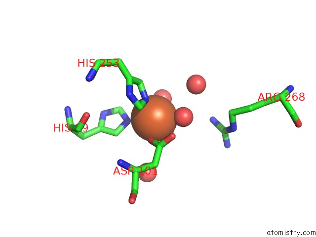
Iron binding site 1 out of 2 in 3r1j
Go back to
Iron binding site 1 out
of 2 in the Crystal Structure of Alpha-Ketoglutarate-Dependent Taurine Dioxygenase From Mycobacterium Avium, Native Form
Mono view

Stereo pair view
Mono view
Stereo pair view
A full contact list of Iron with other atoms in the Fe binding
site number 1 of Crystal Structure of Alpha-Ketoglutarate-Dependent Taurine Dioxygenase From Mycobacterium Avium, Native Form within 5.0Å range:
|
Iron binding site 2 out of 2 in 3r1j
Go back to
Iron binding site 2 out
of 2 in the Crystal Structure of Alpha-Ketoglutarate-Dependent Taurine Dioxygenase From Mycobacterium Avium, Native Form

Mono view

Stereo pair view
Mono view
Stereo pair view
A full contact list of Iron with other atoms in the Fe binding
site number 2 of Crystal Structure of Alpha-Ketoglutarate-Dependent Taurine Dioxygenase From Mycobacterium Avium, Native Form within 5.0Å range:
|
Reference:
L.Baugh,
I.Phan,
D.W.Begley,
M.C.Clifton,
B.Armour,
D.M.Dranow,
B.M.Taylor,
M.M.Muruthi,
J.Abendroth,
J.W.Fairman,
D.Fox,
S.H.Dieterich,
B.L.Staker,
A.S.Gardberg,
R.Choi,
S.N.Hewitt,
A.J.Napuli,
J.Myers,
L.K.Barrett,
Y.Zhang,
M.Ferrell,
E.Mundt,
K.Thompkins,
N.Tran,
S.Lyons-Abbott,
A.Abramov,
A.Sekar,
D.Serbzhinskiy,
D.Lorimer,
G.W.Buchko,
R.Stacy,
L.J.Stewart,
T.E.Edwards,
W.C.Van Voorhis,
P.J.Myler.
Increasing the Structural Coverage of Tuberculosis Drug Targets. Tuberculosis (Edinb) V. 95 142 2015.
ISSN: ISSN 1472-9792
PubMed: 25613812
DOI: 10.1016/J.TUBE.2014.12.003
Page generated: Tue Aug 5 06:19:08 2025
ISSN: ISSN 1472-9792
PubMed: 25613812
DOI: 10.1016/J.TUBE.2014.12.003
Last articles
I in 5IJQI in 5JRV
I in 5JGP
I in 5IO8
I in 5IJS
I in 5IJW
I in 5GZH
I in 5IE1
I in 5I3W
I in 5HOW