Iron »
PDB 3r1a-3rmk »
3rfa »
Iron in PDB 3rfa: X-Ray Structure of Rlmn From Escherichia Coli in Complex with S- Adenosylmethionine
Enzymatic activity of X-Ray Structure of Rlmn From Escherichia Coli in Complex with S- Adenosylmethionine
All present enzymatic activity of X-Ray Structure of Rlmn From Escherichia Coli in Complex with S- Adenosylmethionine:
2.1.1.192;
2.1.1.192;
Protein crystallography data
The structure of X-Ray Structure of Rlmn From Escherichia Coli in Complex with S- Adenosylmethionine, PDB code: 3rfa
was solved by
A.K.Boal,
T.L.Grove,
M.I.Mclaughlin,
N.Yennawar,
S.J.Booker,
A.C.Rosenzweig,
with X-Ray Crystallography technique. A brief refinement statistics is given in the table below:
| Resolution Low / High (Å) | 30.00 / 2.05 |
| Space group | P 21 21 21 |
| Cell size a, b, c (Å), α, β, γ (°) | 55.180, 55.621, 252.179, 90.00, 90.00, 90.00 |
| R / Rfree (%) | 20.2 / 24.1 |
Iron Binding Sites:
The binding sites of Iron atom in the X-Ray Structure of Rlmn From Escherichia Coli in Complex with S- Adenosylmethionine
(pdb code 3rfa). This binding sites where shown within
5.0 Angstroms radius around Iron atom.
In total 8 binding sites of Iron where determined in the X-Ray Structure of Rlmn From Escherichia Coli in Complex with S- Adenosylmethionine, PDB code: 3rfa:
Jump to Iron binding site number: 1; 2; 3; 4; 5; 6; 7; 8;
In total 8 binding sites of Iron where determined in the X-Ray Structure of Rlmn From Escherichia Coli in Complex with S- Adenosylmethionine, PDB code: 3rfa:
Jump to Iron binding site number: 1; 2; 3; 4; 5; 6; 7; 8;
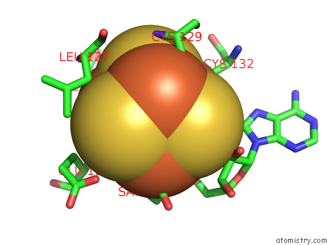
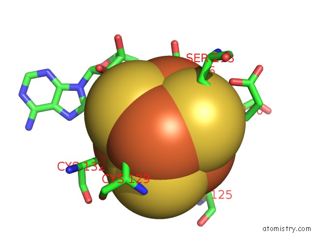
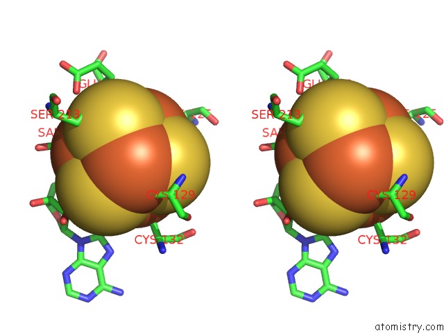
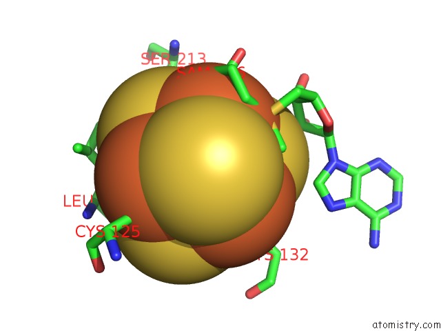
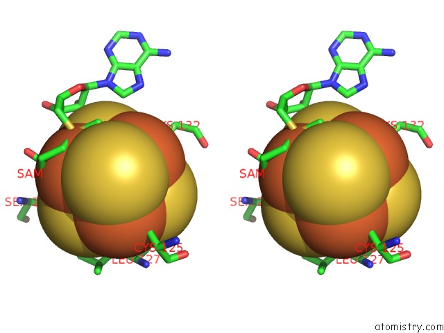
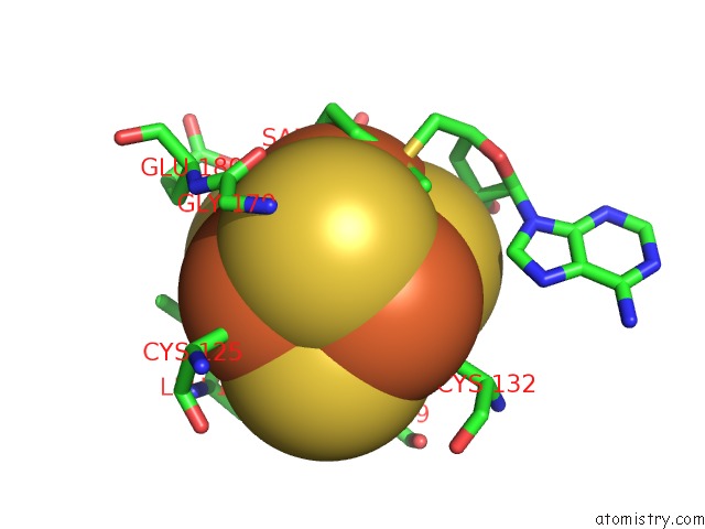
Iron binding site 1 out of 8 in 3rfa
Go back to
Iron binding site 1 out
of 8 in the X-Ray Structure of Rlmn From Escherichia Coli in Complex with S- Adenosylmethionine
Mono view
Stereo pair view
Mono view
Stereo pair view
A full contact list of Iron with other atoms in the Fe binding
site number 1 of X-Ray Structure of Rlmn From Escherichia Coli in Complex with S- Adenosylmethionine within 5.0Å range:
|
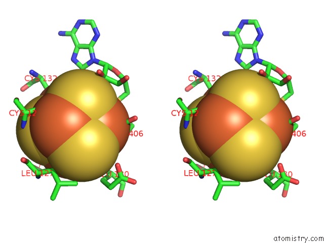
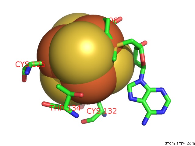
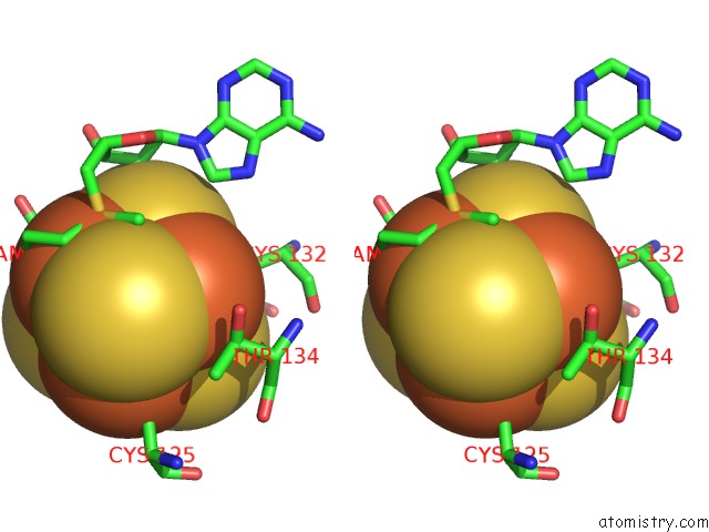
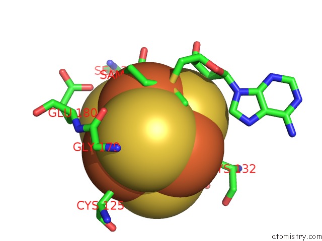
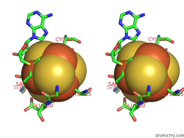
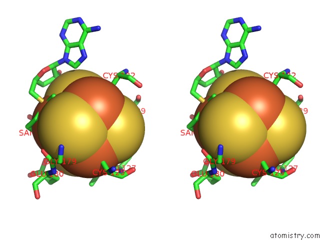
Iron binding site 2 out of 8 in 3rfa
Go back to
Iron binding site 2 out
of 8 in the X-Ray Structure of Rlmn From Escherichia Coli in Complex with S- Adenosylmethionine
Mono view
Stereo pair view
Mono view
Stereo pair view
A full contact list of Iron with other atoms in the Fe binding
site number 2 of X-Ray Structure of Rlmn From Escherichia Coli in Complex with S- Adenosylmethionine within 5.0Å range:
|
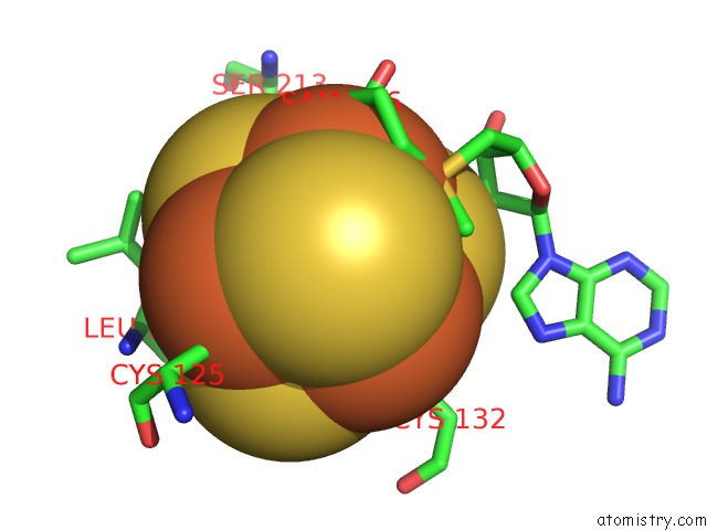
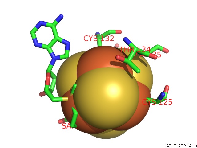
Iron binding site 3 out of 8 in 3rfa
Go back to
Iron binding site 3 out
of 8 in the X-Ray Structure of Rlmn From Escherichia Coli in Complex with S- Adenosylmethionine
Mono view
Stereo pair view
Mono view
Stereo pair view
A full contact list of Iron with other atoms in the Fe binding
site number 3 of X-Ray Structure of Rlmn From Escherichia Coli in Complex with S- Adenosylmethionine within 5.0Å range:
|
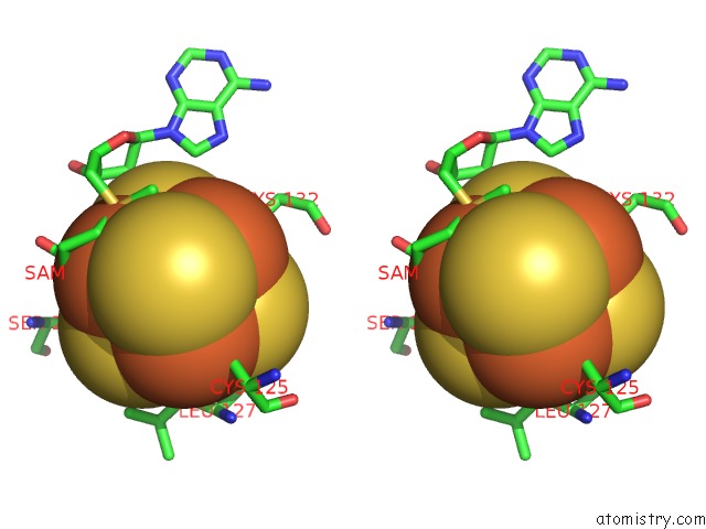
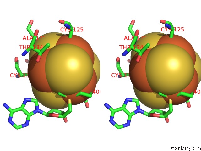
Iron binding site 4 out of 8 in 3rfa
Go back to
Iron binding site 4 out
of 8 in the X-Ray Structure of Rlmn From Escherichia Coli in Complex with S- Adenosylmethionine
Mono view
Stereo pair view
Mono view
Stereo pair view
A full contact list of Iron with other atoms in the Fe binding
site number 4 of X-Ray Structure of Rlmn From Escherichia Coli in Complex with S- Adenosylmethionine within 5.0Å range:
|
Iron binding site 5 out of 8 in 3rfa
Go back to
Iron binding site 5 out
of 8 in the X-Ray Structure of Rlmn From Escherichia Coli in Complex with S- Adenosylmethionine
Mono view
Stereo pair view
Mono view
Stereo pair view
A full contact list of Iron with other atoms in the Fe binding
site number 5 of X-Ray Structure of Rlmn From Escherichia Coli in Complex with S- Adenosylmethionine within 5.0Å range:
|
Iron binding site 6 out of 8 in 3rfa
Go back to
Iron binding site 6 out
of 8 in the X-Ray Structure of Rlmn From Escherichia Coli in Complex with S- Adenosylmethionine
Mono view
Stereo pair view
Mono view
Stereo pair view
A full contact list of Iron with other atoms in the Fe binding
site number 6 of X-Ray Structure of Rlmn From Escherichia Coli in Complex with S- Adenosylmethionine within 5.0Å range:
|
Iron binding site 7 out of 8 in 3rfa
Go back to
Iron binding site 7 out
of 8 in the X-Ray Structure of Rlmn From Escherichia Coli in Complex with S- Adenosylmethionine
Mono view
Stereo pair view
Mono view
Stereo pair view
A full contact list of Iron with other atoms in the Fe binding
site number 7 of X-Ray Structure of Rlmn From Escherichia Coli in Complex with S- Adenosylmethionine within 5.0Å range:
|
Iron binding site 8 out of 8 in 3rfa
Go back to
Iron binding site 8 out
of 8 in the X-Ray Structure of Rlmn From Escherichia Coli in Complex with S- Adenosylmethionine
Mono view
Stereo pair view
Mono view
Stereo pair view
A full contact list of Iron with other atoms in the Fe binding
site number 8 of X-Ray Structure of Rlmn From Escherichia Coli in Complex with S- Adenosylmethionine within 5.0Å range:
|
Reference:
A.K.Boal,
T.L.Grove,
M.I.Mclaughlin,
N.H.Yennawar,
S.J.Booker,
A.C.Rosenzweig.
Structural Basis For Methyl Transfer By A Radical Sam Enzyme. Science V. 332 1089 2011.
ISSN: ISSN 0036-8075
PubMed: 21527678
DOI: 10.1126/SCIENCE.1205358
Page generated: Tue Aug 5 06:23:26 2025
ISSN: ISSN 0036-8075
PubMed: 21527678
DOI: 10.1126/SCIENCE.1205358
Last articles
K in 5W5KK in 5W77
K in 5W8P
K in 5WAY
K in 5W53
K in 5W6P
K in 5W6S
K in 5W6H
K in 5W4M
K in 5W19