Iron »
PDB 3r1a-3rmk »
3rj6 »
Iron in PDB 3rj6: Crystal Structure of Horse Heart Ferric Myoglobin; K45E/K63E/K96E Mutant
Protein crystallography data
The structure of Crystal Structure of Horse Heart Ferric Myoglobin; K45E/K63E/K96E Mutant, PDB code: 3rj6
was solved by
S.M.Smith,
A.C.Rosenzweig,
with X-Ray Crystallography technique. A brief refinement statistics is given in the table below:
| Resolution Low / High (Å) | 61.78 / 1.23 |
| Space group | P 1 21 1 |
| Cell size a, b, c (Å), α, β, γ (°) | 28.341, 123.565, 34.986, 90.00, 89.73, 90.00 |
| R / Rfree (%) | 17.3 / 19.7 |
Iron Binding Sites:
The binding sites of Iron atom in the Crystal Structure of Horse Heart Ferric Myoglobin; K45E/K63E/K96E Mutant
(pdb code 3rj6). This binding sites where shown within
5.0 Angstroms radius around Iron atom.
In total 2 binding sites of Iron where determined in the Crystal Structure of Horse Heart Ferric Myoglobin; K45E/K63E/K96E Mutant, PDB code: 3rj6:
Jump to Iron binding site number: 1; 2;
In total 2 binding sites of Iron where determined in the Crystal Structure of Horse Heart Ferric Myoglobin; K45E/K63E/K96E Mutant, PDB code: 3rj6:
Jump to Iron binding site number: 1; 2;
Iron binding site 1 out of 2 in 3rj6
Go back to
Iron binding site 1 out
of 2 in the Crystal Structure of Horse Heart Ferric Myoglobin; K45E/K63E/K96E Mutant
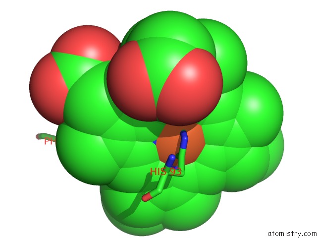
Mono view
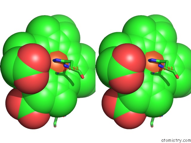
Stereo pair view
Mono view
Stereo pair view
A full contact list of Iron with other atoms in the Fe binding
site number 1 of Crystal Structure of Horse Heart Ferric Myoglobin; K45E/K63E/K96E Mutant within 5.0Å range:
|
Iron binding site 2 out of 2 in 3rj6
Go back to
Iron binding site 2 out
of 2 in the Crystal Structure of Horse Heart Ferric Myoglobin; K45E/K63E/K96E Mutant
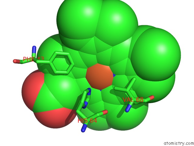
Mono view
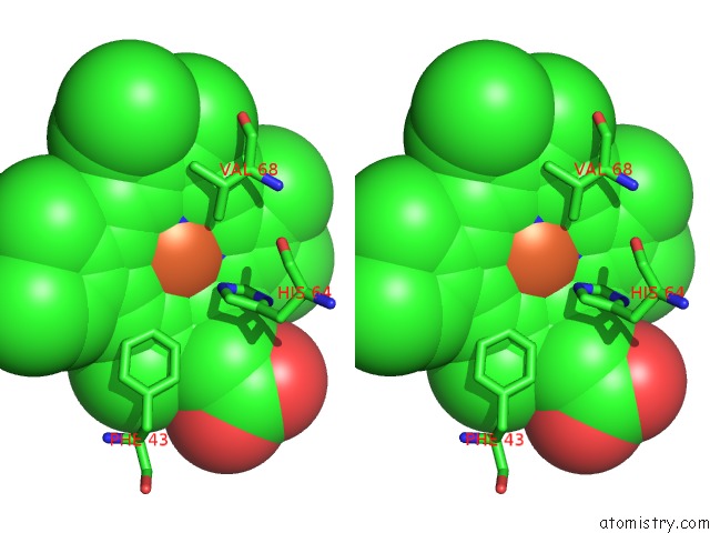
Stereo pair view
Mono view
Stereo pair view
A full contact list of Iron with other atoms in the Fe binding
site number 2 of Crystal Structure of Horse Heart Ferric Myoglobin; K45E/K63E/K96E Mutant within 5.0Å range:
|
Reference:
A.K.Knutson,
S.M.Smith,
A.C.Rosenzweig,
B.M.Hoffman.
Crystal Structure of Horse Heart Ferric Myoglobin; K45E/K63E/K96E Mutant To Be Published.
Page generated: Tue Aug 5 06:29:40 2025
Last articles
K in 2QBLK in 2Q8I
K in 2Q8H
K in 2Q01
K in 2Q8G
K in 2Q8F
K in 2Q3V
K in 2PGA
K in 2PMU
K in 2PUR