Iron »
PDB 3rmz-3s66 »
3ryg »
Iron in PDB 3ryg: 128 Hours Neutron Structure of Perdeuterated Rubredoxin
Iron Binding Sites:
The binding sites of Iron atom in the 128 Hours Neutron Structure of Perdeuterated Rubredoxin
(pdb code 3ryg). This binding sites where shown within
5.0 Angstroms radius around Iron atom.
In total only one binding site of Iron was determined in the 128 Hours Neutron Structure of Perdeuterated Rubredoxin, PDB code: 3ryg:
In total only one binding site of Iron was determined in the 128 Hours Neutron Structure of Perdeuterated Rubredoxin, PDB code: 3ryg:
Iron binding site 1 out of 1 in 3ryg
Go back to
Iron binding site 1 out
of 1 in the 128 Hours Neutron Structure of Perdeuterated Rubredoxin
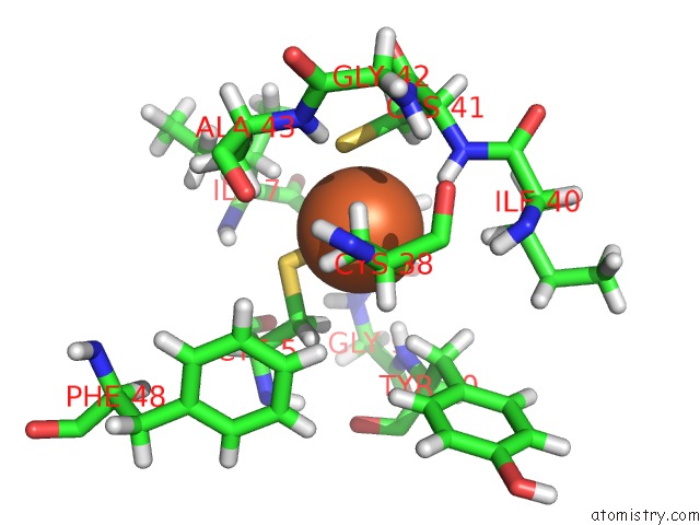
Mono view
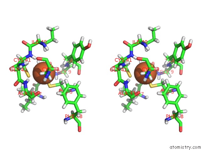
Stereo pair view
Mono view
Stereo pair view
A full contact list of Iron with other atoms in the Fe binding
site number 1 of 128 Hours Neutron Structure of Perdeuterated Rubredoxin within 5.0Å range:
|
Reference:
P.Munshi,
S.L.Chung,
M.P.Blakeley,
K.L.Weiss,
D.A.Myles,
F.Meilleur.
Rapid Visualization of Hydrogen Positions in Protein Neutron Crystallographic Structures. Acta Crystallogr.,Sect.D V. 68 35 2012.
ISSN: ISSN 0907-4449
PubMed: 22194331
DOI: 10.1107/S0907444911048402
Page generated: Tue Aug 5 06:38:21 2025
ISSN: ISSN 0907-4449
PubMed: 22194331
DOI: 10.1107/S0907444911048402
Last articles
Mn in 9LJUMn in 9LJW
Mn in 9LJS
Mn in 9LJR
Mn in 9LJT
Mn in 9LJV
Mg in 9UA2
Mg in 9R96
Mg in 9VM1
Mg in 9P01