Iron »
PDB 3rmz-3s66 »
3s65 »
Iron in PDB 3s65: Structures and Oxygen Affinities of Crystalline Human Hemoglobin C (BETA6 Lys) in the R2 Quaternary Structures
Protein crystallography data
The structure of Structures and Oxygen Affinities of Crystalline Human Hemoglobin C (BETA6 Lys) in the R2 Quaternary Structures, PDB code: 3s65
was solved by
N.Shibayama,
K.Sugiyama,
S.Y.Park,
with X-Ray Crystallography technique. A brief refinement statistics is given in the table below:
| Resolution Low / High (Å) | 19.40 / 1.80 |
| Space group | P 21 21 21 |
| Cell size a, b, c (Å), α, β, γ (°) | 57.784, 58.748, 172.869, 90.00, 90.00, 90.00 |
| R / Rfree (%) | 25 / 30.5 |
Iron Binding Sites:
The binding sites of Iron atom in the Structures and Oxygen Affinities of Crystalline Human Hemoglobin C (BETA6 Lys) in the R2 Quaternary Structures
(pdb code 3s65). This binding sites where shown within
5.0 Angstroms radius around Iron atom.
In total 4 binding sites of Iron where determined in the Structures and Oxygen Affinities of Crystalline Human Hemoglobin C (BETA6 Lys) in the R2 Quaternary Structures, PDB code: 3s65:
Jump to Iron binding site number: 1; 2; 3; 4;
In total 4 binding sites of Iron where determined in the Structures and Oxygen Affinities of Crystalline Human Hemoglobin C (BETA6 Lys) in the R2 Quaternary Structures, PDB code: 3s65:
Jump to Iron binding site number: 1; 2; 3; 4;
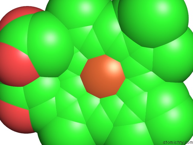
Iron binding site 1 out of 4 in 3s65
Go back to
Iron binding site 1 out
of 4 in the Structures and Oxygen Affinities of Crystalline Human Hemoglobin C (BETA6 Lys) in the R2 Quaternary Structures
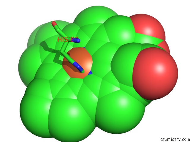
Mono view
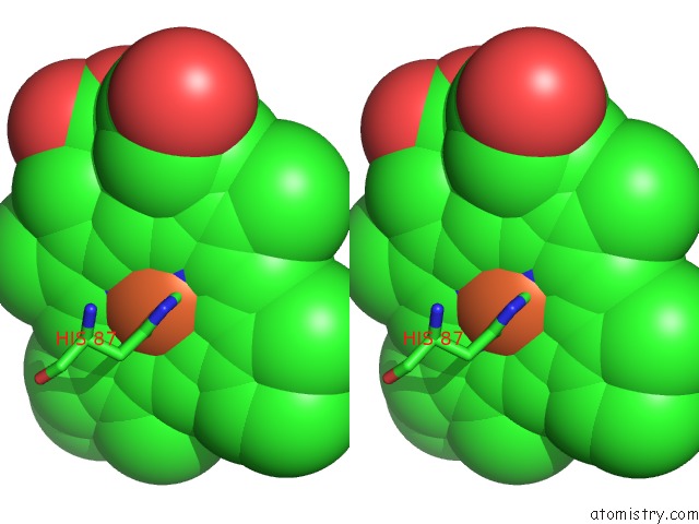
Stereo pair view
Mono view
Stereo pair view
A full contact list of Iron with other atoms in the Fe binding
site number 1 of Structures and Oxygen Affinities of Crystalline Human Hemoglobin C (BETA6 Lys) in the R2 Quaternary Structures within 5.0Å range:
|
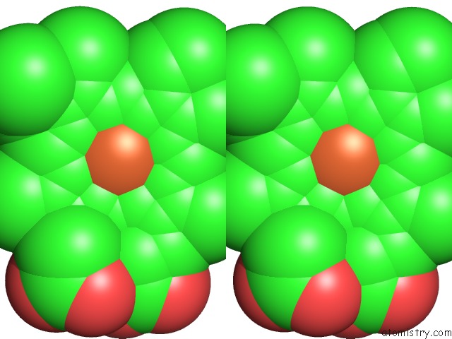
Iron binding site 2 out of 4 in 3s65
Go back to
Iron binding site 2 out
of 4 in the Structures and Oxygen Affinities of Crystalline Human Hemoglobin C (BETA6 Lys) in the R2 Quaternary Structures
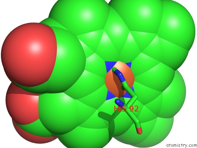
Mono view
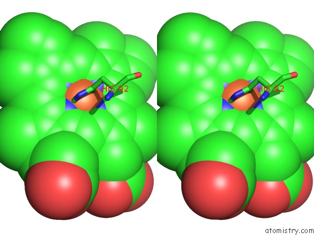
Stereo pair view
Mono view
Stereo pair view
A full contact list of Iron with other atoms in the Fe binding
site number 2 of Structures and Oxygen Affinities of Crystalline Human Hemoglobin C (BETA6 Lys) in the R2 Quaternary Structures within 5.0Å range:
|
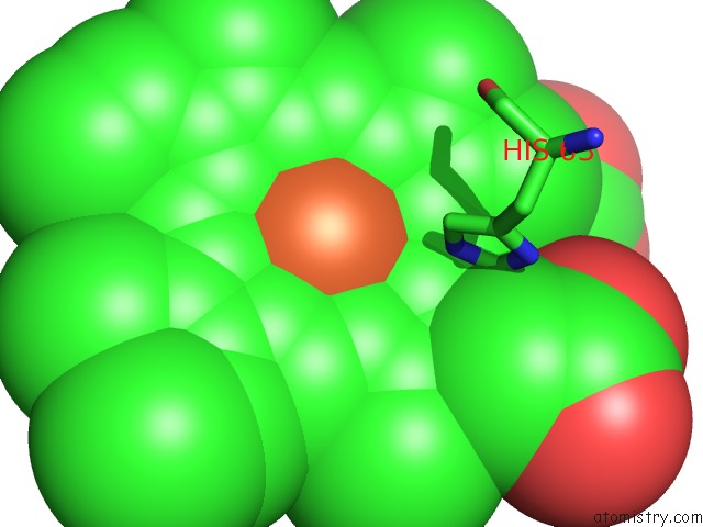
Iron binding site 3 out of 4 in 3s65
Go back to
Iron binding site 3 out
of 4 in the Structures and Oxygen Affinities of Crystalline Human Hemoglobin C (BETA6 Lys) in the R2 Quaternary Structures
Mono view
Stereo pair view
Mono view
Stereo pair view
A full contact list of Iron with other atoms in the Fe binding
site number 3 of Structures and Oxygen Affinities of Crystalline Human Hemoglobin C (BETA6 Lys) in the R2 Quaternary Structures within 5.0Å range:
|
Iron binding site 4 out of 4 in 3s65
Go back to
Iron binding site 4 out
of 4 in the Structures and Oxygen Affinities of Crystalline Human Hemoglobin C (BETA6 Lys) in the R2 Quaternary Structures
Mono view
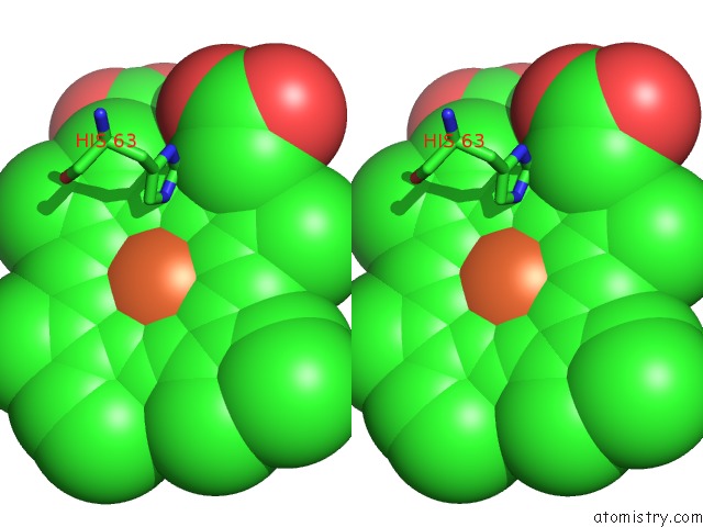
Stereo pair view
Mono view
Stereo pair view
A full contact list of Iron with other atoms in the Fe binding
site number 4 of Structures and Oxygen Affinities of Crystalline Human Hemoglobin C (BETA6 Lys) in the R2 Quaternary Structures within 5.0Å range:
|
Reference:
N.Shibayama,
K.Sugiyama,
S.Y.Park.
Structures and Oxygen Affinities of Crystalline Human Hemoglobin C (BETA6 Glu->Lys) in the R and R2 Quaternary Structure To Be Published.
Page generated: Tue Aug 5 06:40:40 2025
Last articles
Mn in 9LJUMn in 9LJW
Mn in 9LJS
Mn in 9LJR
Mn in 9LJT
Mn in 9LJV
Mg in 9UA2
Mg in 9R96
Mg in 9VM1
Mg in 9P01