Iron »
PDB 3s6b-3sxt »
3sjl »
Iron in PDB 3sjl: Crystal Structure of the P107S-Maug/Pre-Methylamine Dehydrogenase Complex
Enzymatic activity of Crystal Structure of the P107S-Maug/Pre-Methylamine Dehydrogenase Complex
All present enzymatic activity of Crystal Structure of the P107S-Maug/Pre-Methylamine Dehydrogenase Complex:
1.4.99.3;
1.4.99.3;
Protein crystallography data
The structure of Crystal Structure of the P107S-Maug/Pre-Methylamine Dehydrogenase Complex, PDB code: 3sjl
was solved by
L.M.R.Jensen,
C.M.Wilmot,
with X-Ray Crystallography technique. A brief refinement statistics is given in the table below:
| Resolution Low / High (Å) | 29.39 / 1.63 |
| Space group | P 1 |
| Cell size a, b, c (Å), α, β, γ (°) | 55.606, 89.001, 104.812, 67.05, 79.51, 79.72 |
| R / Rfree (%) | 14.2 / 18 |
Other elements in 3sjl:
The structure of Crystal Structure of the P107S-Maug/Pre-Methylamine Dehydrogenase Complex also contains other interesting chemical elements:
| Calcium | (Ca) | 2 atoms |
| Sodium | (Na) | 4 atoms |
Iron Binding Sites:
The binding sites of Iron atom in the Crystal Structure of the P107S-Maug/Pre-Methylamine Dehydrogenase Complex
(pdb code 3sjl). This binding sites where shown within
5.0 Angstroms radius around Iron atom.
In total 4 binding sites of Iron where determined in the Crystal Structure of the P107S-Maug/Pre-Methylamine Dehydrogenase Complex, PDB code: 3sjl:
Jump to Iron binding site number: 1; 2; 3; 4;
In total 4 binding sites of Iron where determined in the Crystal Structure of the P107S-Maug/Pre-Methylamine Dehydrogenase Complex, PDB code: 3sjl:
Jump to Iron binding site number: 1; 2; 3; 4;
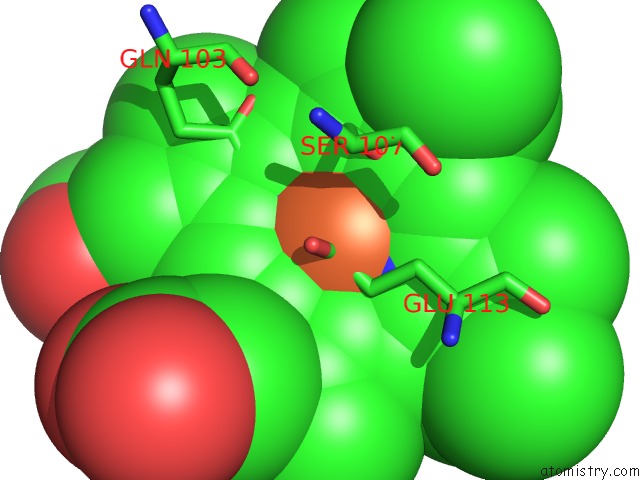
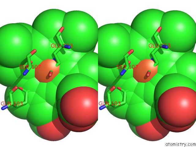
Iron binding site 1 out of 4 in 3sjl
Go back to
Iron binding site 1 out
of 4 in the Crystal Structure of the P107S-Maug/Pre-Methylamine Dehydrogenase Complex
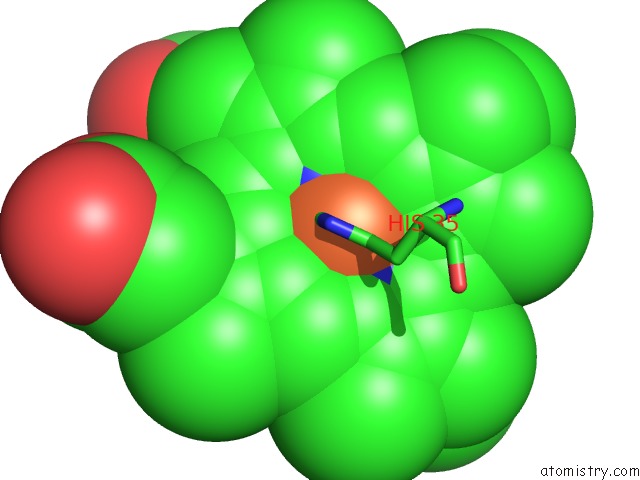
Mono view
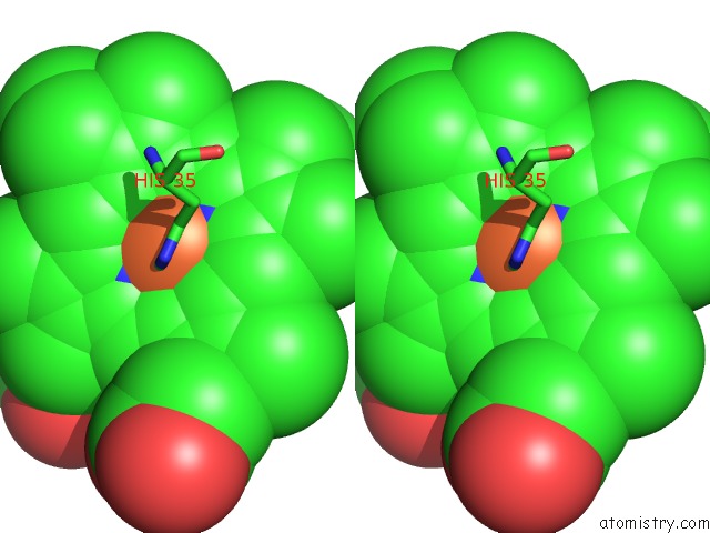
Stereo pair view
Mono view
Stereo pair view
A full contact list of Iron with other atoms in the Fe binding
site number 1 of Crystal Structure of the P107S-Maug/Pre-Methylamine Dehydrogenase Complex within 5.0Å range:
|
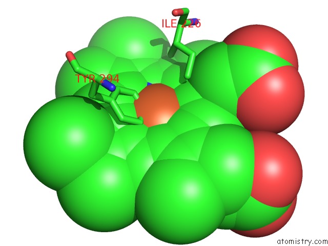
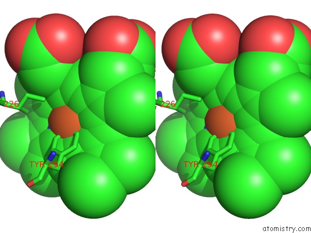
Iron binding site 2 out of 4 in 3sjl
Go back to
Iron binding site 2 out
of 4 in the Crystal Structure of the P107S-Maug/Pre-Methylamine Dehydrogenase Complex
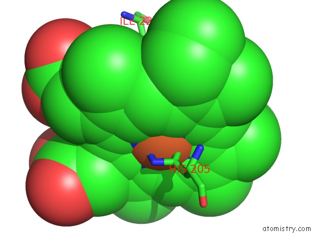
Mono view
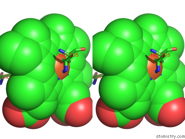
Stereo pair view
Mono view
Stereo pair view
A full contact list of Iron with other atoms in the Fe binding
site number 2 of Crystal Structure of the P107S-Maug/Pre-Methylamine Dehydrogenase Complex within 5.0Å range:
|
Iron binding site 3 out of 4 in 3sjl
Go back to
Iron binding site 3 out
of 4 in the Crystal Structure of the P107S-Maug/Pre-Methylamine Dehydrogenase Complex
Mono view
Stereo pair view
Mono view
Stereo pair view
A full contact list of Iron with other atoms in the Fe binding
site number 3 of Crystal Structure of the P107S-Maug/Pre-Methylamine Dehydrogenase Complex within 5.0Å range:
|
Iron binding site 4 out of 4 in 3sjl
Go back to
Iron binding site 4 out
of 4 in the Crystal Structure of the P107S-Maug/Pre-Methylamine Dehydrogenase Complex
Mono view
Stereo pair view
Mono view
Stereo pair view
A full contact list of Iron with other atoms in the Fe binding
site number 4 of Crystal Structure of the P107S-Maug/Pre-Methylamine Dehydrogenase Complex within 5.0Å range:
|
Reference:
M.Feng,
L.M.Jensen,
E.T.Yukl,
X.Wei,
A.Liu,
C.M.Wilmot,
V.L.Davidson.
Proline 107 Is A Major Determinant in Maintaining the Structure of the Distal Pocket and Reactivity of the High-Spin Heme of Maug. Biochemistry V. 51 1598 2012.
ISSN: ISSN 0006-2960
PubMed: 22299652
DOI: 10.1021/BI201882E
Page generated: Tue Aug 5 06:48:20 2025
ISSN: ISSN 0006-2960
PubMed: 22299652
DOI: 10.1021/BI201882E
Last articles
K in 1M6VK in 1N31
K in 1N2T
K in 1N0H
K in 1N0X
K in 1MXC
K in 1MXB
K in 1MPW
K in 1MXA
K in 1MP0