Iron »
PDB 3sxv-3tgm »
3tgm »
Iron in PDB 3tgm: X-Ray Crystal Structure of Human Heme Oxygenase-1 in Complex with 1- (1H-Imidazol-1-Yl)-4,4-Diphenyl-2 Butanone
Enzymatic activity of X-Ray Crystal Structure of Human Heme Oxygenase-1 in Complex with 1- (1H-Imidazol-1-Yl)-4,4-Diphenyl-2 Butanone
All present enzymatic activity of X-Ray Crystal Structure of Human Heme Oxygenase-1 in Complex with 1- (1H-Imidazol-1-Yl)-4,4-Diphenyl-2 Butanone:
1.14.99.3;
1.14.99.3;
Protein crystallography data
The structure of X-Ray Crystal Structure of Human Heme Oxygenase-1 in Complex with 1- (1H-Imidazol-1-Yl)-4,4-Diphenyl-2 Butanone, PDB code: 3tgm
was solved by
M.N.Rahman,
Z.Jia,
with X-Ray Crystallography technique. A brief refinement statistics is given in the table below:
| Resolution Low / High (Å) | 19.82 / 2.85 |
| Space group | P 21 21 21 |
| Cell size a, b, c (Å), α, β, γ (°) | 54.610, 74.980, 115.280, 90.00, 90.00, 90.00 |
| R / Rfree (%) | 22.3 / 26.4 |
Iron Binding Sites:
The binding sites of Iron atom in the X-Ray Crystal Structure of Human Heme Oxygenase-1 in Complex with 1- (1H-Imidazol-1-Yl)-4,4-Diphenyl-2 Butanone
(pdb code 3tgm). This binding sites where shown within
5.0 Angstroms radius around Iron atom.
In total 2 binding sites of Iron where determined in the X-Ray Crystal Structure of Human Heme Oxygenase-1 in Complex with 1- (1H-Imidazol-1-Yl)-4,4-Diphenyl-2 Butanone, PDB code: 3tgm:
Jump to Iron binding site number: 1; 2;
In total 2 binding sites of Iron where determined in the X-Ray Crystal Structure of Human Heme Oxygenase-1 in Complex with 1- (1H-Imidazol-1-Yl)-4,4-Diphenyl-2 Butanone, PDB code: 3tgm:
Jump to Iron binding site number: 1; 2;
Iron binding site 1 out of 2 in 3tgm
Go back to
Iron binding site 1 out
of 2 in the X-Ray Crystal Structure of Human Heme Oxygenase-1 in Complex with 1- (1H-Imidazol-1-Yl)-4,4-Diphenyl-2 Butanone
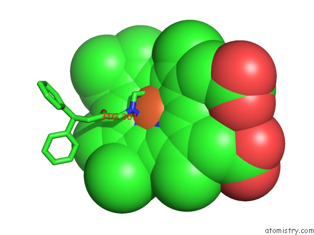
Mono view
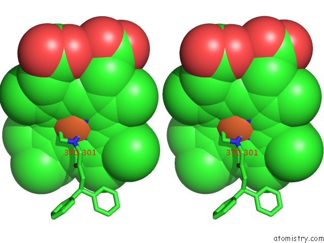
Stereo pair view
Mono view
Stereo pair view
A full contact list of Iron with other atoms in the Fe binding
site number 1 of X-Ray Crystal Structure of Human Heme Oxygenase-1 in Complex with 1- (1H-Imidazol-1-Yl)-4,4-Diphenyl-2 Butanone within 5.0Å range:
|
Iron binding site 2 out of 2 in 3tgm
Go back to
Iron binding site 2 out
of 2 in the X-Ray Crystal Structure of Human Heme Oxygenase-1 in Complex with 1- (1H-Imidazol-1-Yl)-4,4-Diphenyl-2 Butanone
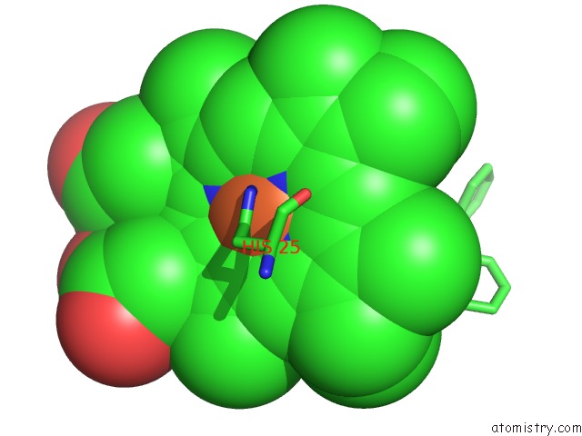
Mono view
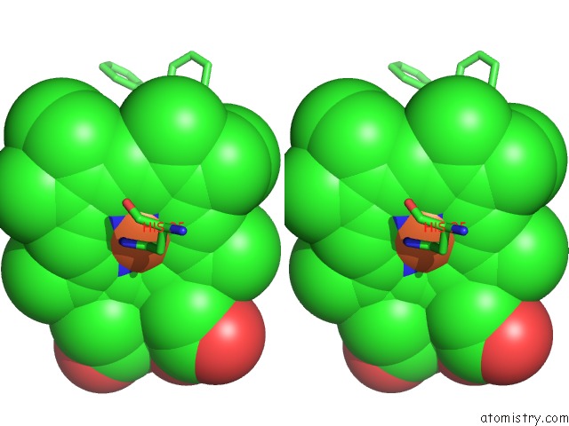
Stereo pair view
Mono view
Stereo pair view
A full contact list of Iron with other atoms in the Fe binding
site number 2 of X-Ray Crystal Structure of Human Heme Oxygenase-1 in Complex with 1- (1H-Imidazol-1-Yl)-4,4-Diphenyl-2 Butanone within 5.0Å range:
|
Reference:
M.N.Rahman,
J.Z.Vlahakis,
D.Vukomanovic,
W.Lee,
W.A.Szarek,
K.Nakatsu,
Z.Jia.
A Novel, "Double-Clamp" Binding Mode For Human Heme Oxygenase-1 Inhibition. Plos One V. 7 29514 2012.
ISSN: ESSN 1932-6203
PubMed: 22276118
DOI: 10.1371/JOURNAL.PONE.0029514
Page generated: Tue Aug 5 07:02:54 2025
ISSN: ESSN 1932-6203
PubMed: 22276118
DOI: 10.1371/JOURNAL.PONE.0029514
Last articles
Na in 3CCONa in 3CC2
Na in 3CCE
Na in 3CC7
Na in 3CC4
Na in 3CC9
Na in 3CBC
Na in 3CBT
Na in 3C9F
Na in 3CB8