Iron »
PDB 3tgu-3u3e »
3tjt »
Iron in PDB 3tjt: Crystal Structure Analysis of the Superoxide Dismutase From Clostridium Difficile
Enzymatic activity of Crystal Structure Analysis of the Superoxide Dismutase From Clostridium Difficile
All present enzymatic activity of Crystal Structure Analysis of the Superoxide Dismutase From Clostridium Difficile:
1.15.1.1;
1.15.1.1;
Protein crystallography data
The structure of Crystal Structure Analysis of the Superoxide Dismutase From Clostridium Difficile, PDB code: 3tjt
was solved by
W.Li,
C.Lei,
T.L.Ying,
H.F.Wang,
X.S.Tan,
with X-Ray Crystallography technique. A brief refinement statistics is given in the table below:
| Resolution Low / High (Å) | 40.79 / 1.80 |
| Space group | P 65 2 2 |
| Cell size a, b, c (Å), α, β, γ (°) | 80.399, 80.399, 251.631, 90.00, 90.00, 120.00 |
| R / Rfree (%) | 18 / 20.3 |
Iron Binding Sites:
The binding sites of Iron atom in the Crystal Structure Analysis of the Superoxide Dismutase From Clostridium Difficile
(pdb code 3tjt). This binding sites where shown within
5.0 Angstroms radius around Iron atom.
In total only one binding site of Iron was determined in the Crystal Structure Analysis of the Superoxide Dismutase From Clostridium Difficile, PDB code: 3tjt:
In total only one binding site of Iron was determined in the Crystal Structure Analysis of the Superoxide Dismutase From Clostridium Difficile, PDB code: 3tjt:
Iron binding site 1 out of 1 in 3tjt
Go back to
Iron binding site 1 out
of 1 in the Crystal Structure Analysis of the Superoxide Dismutase From Clostridium Difficile
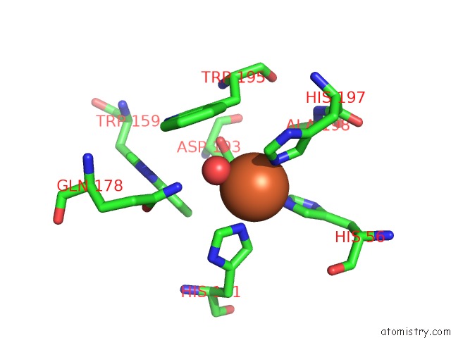
Mono view
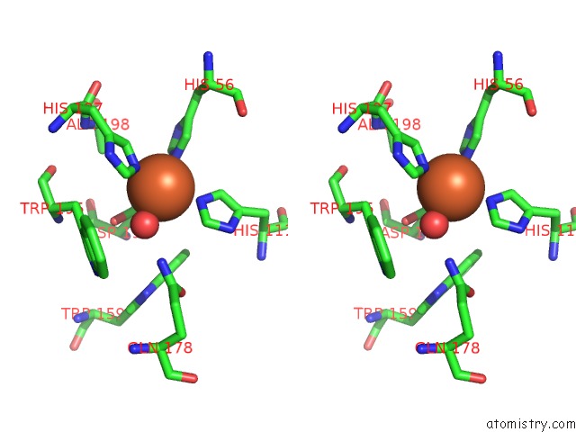
Stereo pair view
Mono view
Stereo pair view
A full contact list of Iron with other atoms in the Fe binding
site number 1 of Crystal Structure Analysis of the Superoxide Dismutase From Clostridium Difficile within 5.0Å range:
|
Reference:
W.Li,
C.Lei,
T.L.Ying,
H.F.Wang,
X.S.Tan.
Crystal Structure of the Superoxide Dismutase From Clostridium Difficile To Be Published.
Page generated: Tue Aug 5 07:03:43 2025
Last articles
Mn in 7NQEMn in 7N8O
Mn in 7NRO
Mn in 7NHQ
Mn in 7NHP
Mn in 7NHO
Mn in 7NF4
Mn in 7NF2
Mn in 7NF3
Mn in 7NF1