Iron »
PDB 3tgu-3u3e »
3tny »
Iron in PDB 3tny: Structure of Yfiy From Bacillus Cereus Bound to the Siderophore Iron (III) Schizokinen
Protein crystallography data
The structure of Structure of Yfiy From Bacillus Cereus Bound to the Siderophore Iron (III) Schizokinen, PDB code: 3tny
was solved by
M.C.Clifton,
with X-Ray Crystallography technique. A brief refinement statistics is given in the table below:
| Resolution Low / High (Å) | 50.00 / 1.55 |
| Space group | P 1 21 1 |
| Cell size a, b, c (Å), α, β, γ (°) | 57.796, 47.328, 64.981, 90.00, 112.88, 90.00 |
| R / Rfree (%) | 17.2 / 18.9 |
Iron Binding Sites:
The binding sites of Iron atom in the Structure of Yfiy From Bacillus Cereus Bound to the Siderophore Iron (III) Schizokinen
(pdb code 3tny). This binding sites where shown within
5.0 Angstroms radius around Iron atom.
In total only one binding site of Iron was determined in the Structure of Yfiy From Bacillus Cereus Bound to the Siderophore Iron (III) Schizokinen, PDB code: 3tny:
In total only one binding site of Iron was determined in the Structure of Yfiy From Bacillus Cereus Bound to the Siderophore Iron (III) Schizokinen, PDB code: 3tny:
Iron binding site 1 out of 1 in 3tny
Go back to
Iron binding site 1 out
of 1 in the Structure of Yfiy From Bacillus Cereus Bound to the Siderophore Iron (III) Schizokinen
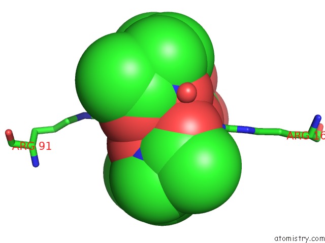
Mono view
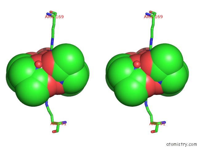
Stereo pair view
Mono view
Stereo pair view
A full contact list of Iron with other atoms in the Fe binding
site number 1 of Structure of Yfiy From Bacillus Cereus Bound to the Siderophore Iron (III) Schizokinen within 5.0Å range:
|
Reference:
M.C.Clifton,
P.B.Rupert,
T.M.Hoette,
K.N.Raymond,
R.J.Abergel,
R.K.Strong.
Parsing the Functional Specificity of Siderocalin / Lipocalin 2 / Ngal For Siderophores and Related Small-Molecule Ligands To Be Published.
Page generated: Tue Aug 5 07:06:32 2025
Last articles
Mn in 7QWJMn in 7QOE
Mn in 7QOD
Mn in 7QHO
Mn in 7QHM
Mn in 7QJJ
Mn in 7QFB
Mn in 7PI0
Mn in 7QBP
Mn in 7QBK