Iron »
PDB 3tgu-3u3e »
3tus »
Iron in PDB 3tus: Crystal Structure of C-Lobe of Bovine Lactoferrin Complexed with Meta- Hydroxy Benzoic Acid at 2.5 A Resolution
Protein crystallography data
The structure of Crystal Structure of C-Lobe of Bovine Lactoferrin Complexed with Meta- Hydroxy Benzoic Acid at 2.5 A Resolution, PDB code: 3tus
was solved by
P.K.Shukla,
L.Gautam,
A.Singh,
S.Kaushik,
M.Sinha,
A.Bhushan,
P.Kaur,
S.Sharma,
T.P.Singh,
with X-Ray Crystallography technique. A brief refinement statistics is given in the table below:
| Resolution Low / High (Å) | 39.26 / 2.50 |
| Space group | P 1 21 1 |
| Cell size a, b, c (Å), α, β, γ (°) | 62.431, 50.492, 65.404, 90.00, 107.32, 90.00 |
| R / Rfree (%) | 21.8 / 24.1 |
Other elements in 3tus:
The structure of Crystal Structure of C-Lobe of Bovine Lactoferrin Complexed with Meta- Hydroxy Benzoic Acid at 2.5 A Resolution also contains other interesting chemical elements:
| Zinc | (Zn) | 2 atoms |
Iron Binding Sites:
The binding sites of Iron atom in the Crystal Structure of C-Lobe of Bovine Lactoferrin Complexed with Meta- Hydroxy Benzoic Acid at 2.5 A Resolution
(pdb code 3tus). This binding sites where shown within
5.0 Angstroms radius around Iron atom.
In total only one binding site of Iron was determined in the Crystal Structure of C-Lobe of Bovine Lactoferrin Complexed with Meta- Hydroxy Benzoic Acid at 2.5 A Resolution, PDB code: 3tus:
In total only one binding site of Iron was determined in the Crystal Structure of C-Lobe of Bovine Lactoferrin Complexed with Meta- Hydroxy Benzoic Acid at 2.5 A Resolution, PDB code: 3tus:
Iron binding site 1 out of 1 in 3tus
Go back to
Iron binding site 1 out
of 1 in the Crystal Structure of C-Lobe of Bovine Lactoferrin Complexed with Meta- Hydroxy Benzoic Acid at 2.5 A Resolution
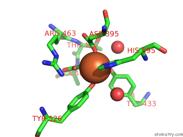
Mono view
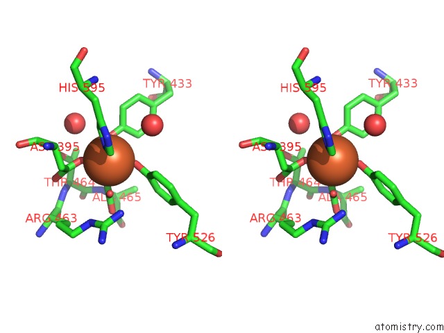
Stereo pair view
Mono view
Stereo pair view
A full contact list of Iron with other atoms in the Fe binding
site number 1 of Crystal Structure of C-Lobe of Bovine Lactoferrin Complexed with Meta- Hydroxy Benzoic Acid at 2.5 A Resolution within 5.0Å range:
|
Reference:
P.K.Shukla,
L.Gautam,
A.Singh,
S.Kaushik,
M.Sinha,
A.Bhushan,
P.Kaur,
S.Sharma,
T.P.Singh.
Crystal Structure of C-Lobe of Bovine Lactoferrin Complexed with Meta-Hydroxy Benzoic Acid at 2.5 A Resolution To Be Published.
Page generated: Tue Aug 5 07:10:35 2025
Last articles
Mn in 7THMMn in 7TDB
Mn in 7T0Y
Mn in 7TD7
Mn in 7TCL
Mn in 7SS7
Mn in 7SS6
Mn in 7SD0
Mn in 7SP8
Mn in 7SP7