Iron »
PDB 3tgu-3u3e »
3u0d »
Iron in PDB 3u0d: The Structure of Human Siderocalin Bound to the Bacterial Siderophore 2,3-Dhba
Protein crystallography data
The structure of The Structure of Human Siderocalin Bound to the Bacterial Siderophore 2,3-Dhba, PDB code: 3u0d
was solved by
Seattle Structural Genomics Center For Infectious Disease (Ssgcid),
with X-Ray Crystallography technique. A brief refinement statistics is given in the table below:
| Resolution Low / High (Å) | 42.01 / 2.51 |
| Space group | P 21 21 21 |
| Cell size a, b, c (Å), α, β, γ (°) | 117.476, 116.452, 120.194, 90.00, 90.00, 90.00 |
| R / Rfree (%) | 24.7 / 28.8 |
Other elements in 3u0d:
The structure of The Structure of Human Siderocalin Bound to the Bacterial Siderophore 2,3-Dhba also contains other interesting chemical elements:
| Chlorine | (Cl) | 3 atoms |
Iron Binding Sites:
The binding sites of Iron atom in the The Structure of Human Siderocalin Bound to the Bacterial Siderophore 2,3-Dhba
(pdb code 3u0d). This binding sites where shown within
5.0 Angstroms radius around Iron atom.
In total 4 binding sites of Iron where determined in the The Structure of Human Siderocalin Bound to the Bacterial Siderophore 2,3-Dhba, PDB code: 3u0d:
Jump to Iron binding site number: 1; 2; 3; 4;
In total 4 binding sites of Iron where determined in the The Structure of Human Siderocalin Bound to the Bacterial Siderophore 2,3-Dhba, PDB code: 3u0d:
Jump to Iron binding site number: 1; 2; 3; 4;
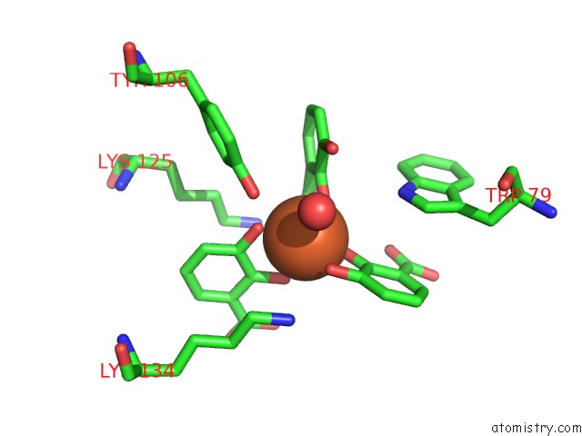
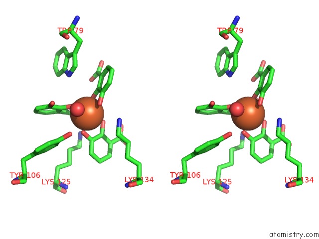
Iron binding site 1 out of 4 in 3u0d
Go back to
Iron binding site 1 out
of 4 in the The Structure of Human Siderocalin Bound to the Bacterial Siderophore 2,3-Dhba
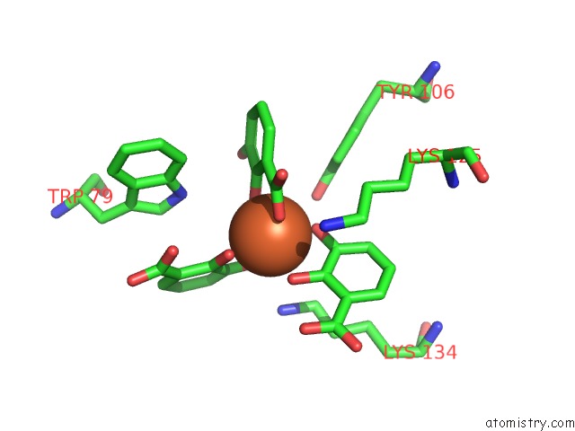
Mono view
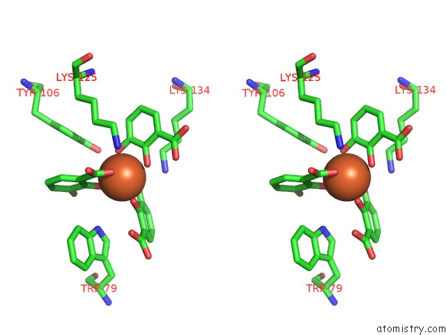
Stereo pair view
Mono view
Stereo pair view
A full contact list of Iron with other atoms in the Fe binding
site number 1 of The Structure of Human Siderocalin Bound to the Bacterial Siderophore 2,3-Dhba within 5.0Å range:
|
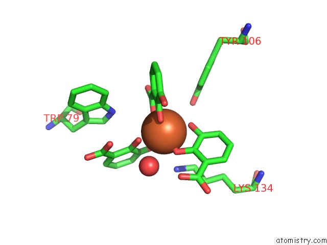
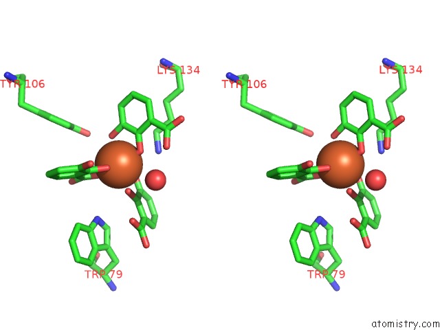
Iron binding site 2 out of 4 in 3u0d
Go back to
Iron binding site 2 out
of 4 in the The Structure of Human Siderocalin Bound to the Bacterial Siderophore 2,3-Dhba
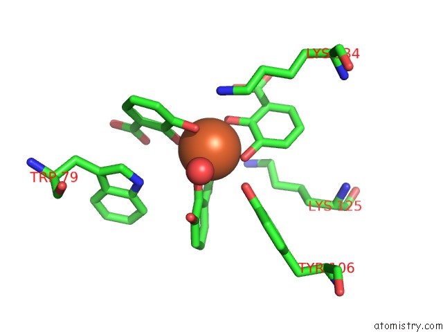
Mono view
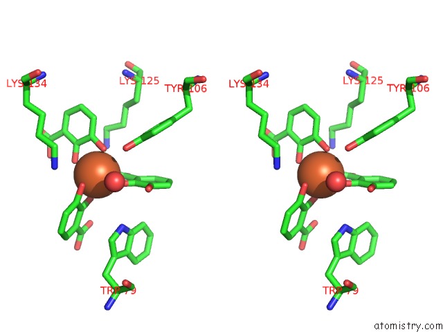
Stereo pair view
Mono view
Stereo pair view
A full contact list of Iron with other atoms in the Fe binding
site number 2 of The Structure of Human Siderocalin Bound to the Bacterial Siderophore 2,3-Dhba within 5.0Å range:
|
Iron binding site 3 out of 4 in 3u0d
Go back to
Iron binding site 3 out
of 4 in the The Structure of Human Siderocalin Bound to the Bacterial Siderophore 2,3-Dhba
Mono view
Stereo pair view
Mono view
Stereo pair view
A full contact list of Iron with other atoms in the Fe binding
site number 3 of The Structure of Human Siderocalin Bound to the Bacterial Siderophore 2,3-Dhba within 5.0Å range:
|
Iron binding site 4 out of 4 in 3u0d
Go back to
Iron binding site 4 out
of 4 in the The Structure of Human Siderocalin Bound to the Bacterial Siderophore 2,3-Dhba
Mono view
Stereo pair view
Mono view
Stereo pair view
A full contact list of Iron with other atoms in the Fe binding
site number 4 of The Structure of Human Siderocalin Bound to the Bacterial Siderophore 2,3-Dhba within 5.0Å range:
|
Reference:
C.Correnti,
V.Richardson,
A.K.Sia,
A.D.Bandaranayake,
M.Ruiz,
Y.Suryo Rahmanto,
Z.Kovacevic,
M.C.Clifton,
M.A.Holmes,
B.K.Kaiser,
J.Barasch,
K.N.Raymond,
D.R.Richardson,
R.K.Strong.
Siderocalin/LCN2/Ngal/24P3 Does Not Drive Apoptosis Through Gentisic Acid Mediated Iron Withdrawal in Hematopoietic Cell Lines. Plos One V. 7 43696 2012.
ISSN: ESSN 1932-6203
PubMed: 22928018
DOI: 10.1371/JOURNAL.PONE.0043696
Page generated: Tue Aug 5 07:12:18 2025
ISSN: ESSN 1932-6203
PubMed: 22928018
DOI: 10.1371/JOURNAL.PONE.0043696
Last articles
Mg in 1KZUMg in 1L2X
Mg in 1L3P
Mg in 1L3J
Mg in 1L2O
Mg in 1L2E
Mg in 1L0O
Mg in 1L1R
Mg in 1KXG
Mg in 1KYR