Iron »
PDB 3v3y-3vks »
3vcp »
Iron in PDB 3vcp: The 2.2 Angstrom Structure of STC2 with Proline Bound in the Active Site
Protein crystallography data
The structure of The 2.2 Angstrom Structure of STC2 with Proline Bound in the Active Site, PDB code: 3vcp
was solved by
K.D.Daughtry,
Y.Xiao,
D.Stoner-Ma,
E.Cho,
A.M.Orville,
P.Liu,
K.N.Allen,
with X-Ray Crystallography technique. A brief refinement statistics is given in the table below:
| Resolution Low / High (Å) | 38.11 / 2.20 |
| Space group | P 63 2 2 |
| Cell size a, b, c (Å), α, β, γ (°) | 97.140, 97.140, 180.080, 90.00, 90.00, 120.00 |
| R / Rfree (%) | 18.7 / 24.2 |
Iron Binding Sites:
The binding sites of Iron atom in the The 2.2 Angstrom Structure of STC2 with Proline Bound in the Active Site
(pdb code 3vcp). This binding sites where shown within
5.0 Angstroms radius around Iron atom.
In total 3 binding sites of Iron where determined in the The 2.2 Angstrom Structure of STC2 with Proline Bound in the Active Site, PDB code: 3vcp:
Jump to Iron binding site number: 1; 2; 3;
In total 3 binding sites of Iron where determined in the The 2.2 Angstrom Structure of STC2 with Proline Bound in the Active Site, PDB code: 3vcp:
Jump to Iron binding site number: 1; 2; 3;
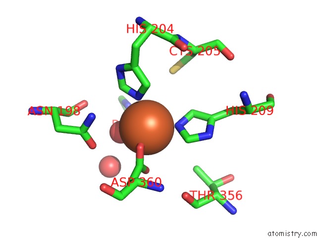
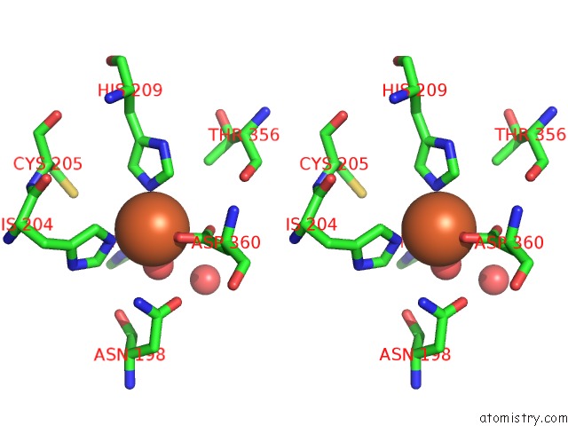
Iron binding site 1 out of 3 in 3vcp
Go back to
Iron binding site 1 out
of 3 in the The 2.2 Angstrom Structure of STC2 with Proline Bound in the Active Site
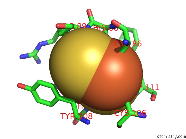
Mono view
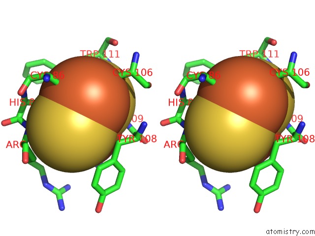
Stereo pair view
Mono view
Stereo pair view
A full contact list of Iron with other atoms in the Fe binding
site number 1 of The 2.2 Angstrom Structure of STC2 with Proline Bound in the Active Site within 5.0Å range:
|
Iron binding site 2 out of 3 in 3vcp
Go back to
Iron binding site 2 out
of 3 in the The 2.2 Angstrom Structure of STC2 with Proline Bound in the Active Site
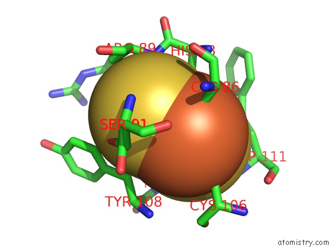
Mono view
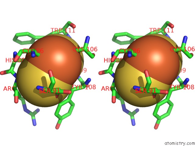
Stereo pair view
Mono view
Stereo pair view
A full contact list of Iron with other atoms in the Fe binding
site number 2 of The 2.2 Angstrom Structure of STC2 with Proline Bound in the Active Site within 5.0Å range:
|
Iron binding site 3 out of 3 in 3vcp
Go back to
Iron binding site 3 out
of 3 in the The 2.2 Angstrom Structure of STC2 with Proline Bound in the Active Site
Mono view
Stereo pair view
Mono view
Stereo pair view
A full contact list of Iron with other atoms in the Fe binding
site number 3 of The 2.2 Angstrom Structure of STC2 with Proline Bound in the Active Site within 5.0Å range:
|
Reference:
K.D.Daughtry,
Y.Xiao,
D.Stoner-Ma,
E.Cho,
A.M.Orville,
P.Liu,
K.N.Allen.
Quaternary Ammonium Oxidative Demethylation: X-Ray Crystallographic, Resonance Raman, and Uv-Visible Spectroscopic Analysis of A Rieske-Type Demethylase. J.Am.Chem.Soc. V. 134 2823 2012.
ISSN: ISSN 0002-7863
PubMed: 22224443
DOI: 10.1021/JA2111898
Page generated: Tue Aug 5 07:51:38 2025
ISSN: ISSN 0002-7863
PubMed: 22224443
DOI: 10.1021/JA2111898
Last articles
Mg in 5SEQMg in 5SEO
Mg in 5SEP
Mg in 5SEN
Mg in 5SEM
Mg in 5SEL
Mg in 5SEK
Mg in 5SEJ
Mg in 5SEI
Mg in 5SEH