Iron »
PDB 3vkt-3vti »
3vnw »
Iron in PDB 3vnw: Crystal Structure of Cytochrome C552 From Thermus Thermophilus at pH 5.44
Protein crystallography data
The structure of Crystal Structure of Cytochrome C552 From Thermus Thermophilus at pH 5.44, PDB code: 3vnw
was solved by
S.Yamada,
N.D.Bouley-Ford,
G.E.Keller,
J.R.Winkler,
H.B.Gray,
with X-Ray Crystallography technique. A brief refinement statistics is given in the table below:
| Resolution Low / High (Å) | 20.00 / 1.97 |
| Space group | P 65 |
| Cell size a, b, c (Å), α, β, γ (°) | 86.980, 86.980, 31.820, 90.00, 90.00, 120.00 |
| R / Rfree (%) | 21.3 / 23.2 |
Iron Binding Sites:
The binding sites of Iron atom in the Crystal Structure of Cytochrome C552 From Thermus Thermophilus at pH 5.44
(pdb code 3vnw). This binding sites where shown within
5.0 Angstroms radius around Iron atom.
In total only one binding site of Iron was determined in the Crystal Structure of Cytochrome C552 From Thermus Thermophilus at pH 5.44, PDB code: 3vnw:
In total only one binding site of Iron was determined in the Crystal Structure of Cytochrome C552 From Thermus Thermophilus at pH 5.44, PDB code: 3vnw:
Iron binding site 1 out of 1 in 3vnw
Go back to
Iron binding site 1 out
of 1 in the Crystal Structure of Cytochrome C552 From Thermus Thermophilus at pH 5.44
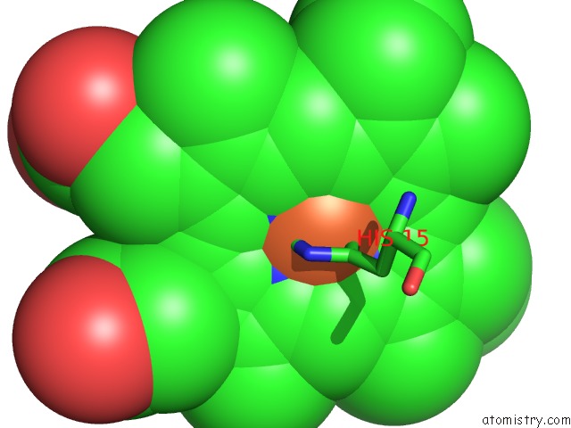
Mono view
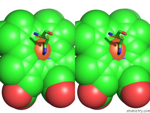
Stereo pair view
Mono view
Stereo pair view
A full contact list of Iron with other atoms in the Fe binding
site number 1 of Crystal Structure of Cytochrome C552 From Thermus Thermophilus at pH 5.44 within 5.0Å range:
|
Reference:
S.Yamada,
N.D.Bouley-Ford,
G.E.Keller,
W.C.Ford,
H.B.Gray,
J.R.Winkler.
Snapshots of A Protein Folding Intermediate Proc.Natl.Acad.Sci.Usa V. 110 1606 2013.
ISSN: ISSN 0027-8424
PubMed: 23319660
DOI: 10.1073/PNAS.1221832110
Page generated: Tue Aug 5 08:02:13 2025
ISSN: ISSN 0027-8424
PubMed: 23319660
DOI: 10.1073/PNAS.1221832110
Last articles
Na in 4O54Na in 4O57
Na in 4O53
Na in 4O50
Na in 4O52
Na in 4O4W
Na in 4O4X
Na in 4O4V
Na in 4O48
Na in 4O47