Iron »
PDB 3vtj-3wg7 »
3wcv »
Iron in PDB 3wcv: The Structure of A Deoxygenated 400 kDa Hemoglobin Provides A More Accurate Description of the Cooperative Mechanism of Giant Hemoglobins: Ca Bound Form
Protein crystallography data
The structure of The Structure of A Deoxygenated 400 kDa Hemoglobin Provides A More Accurate Description of the Cooperative Mechanism of Giant Hemoglobins: Ca Bound Form, PDB code: 3wcv
was solved by
N.Numoto,
T.Nakagawa,
R.Ohara,
T.Hasegawa,
A.Kita,
T.Yoshida,
T.Maruyama,
K.Imai,
Y.Fukumori,
K.Miki,
with X-Ray Crystallography technique. A brief refinement statistics is given in the table below:
| Resolution Low / High (Å) | 36.33 / 2.60 |
| Space group | P 63 |
| Cell size a, b, c (Å), α, β, γ (°) | 108.944, 108.944, 195.016, 90.00, 90.00, 120.00 |
| R / Rfree (%) | 23.5 / 26.7 |
Other elements in 3wcv:
The structure of The Structure of A Deoxygenated 400 kDa Hemoglobin Provides A More Accurate Description of the Cooperative Mechanism of Giant Hemoglobins: Ca Bound Form also contains other interesting chemical elements:
| Calcium | (Ca) | 6 atoms |
Iron Binding Sites:
The binding sites of Iron atom in the The Structure of A Deoxygenated 400 kDa Hemoglobin Provides A More Accurate Description of the Cooperative Mechanism of Giant Hemoglobins: Ca Bound Form
(pdb code 3wcv). This binding sites where shown within
5.0 Angstroms radius around Iron atom.
In total 8 binding sites of Iron where determined in the The Structure of A Deoxygenated 400 kDa Hemoglobin Provides A More Accurate Description of the Cooperative Mechanism of Giant Hemoglobins: Ca Bound Form, PDB code: 3wcv:
Jump to Iron binding site number: 1; 2; 3; 4; 5; 6; 7; 8;
In total 8 binding sites of Iron where determined in the The Structure of A Deoxygenated 400 kDa Hemoglobin Provides A More Accurate Description of the Cooperative Mechanism of Giant Hemoglobins: Ca Bound Form, PDB code: 3wcv:
Jump to Iron binding site number: 1; 2; 3; 4; 5; 6; 7; 8;
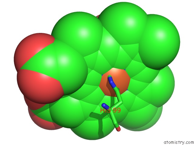
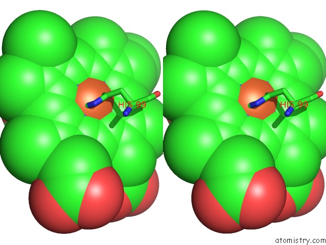
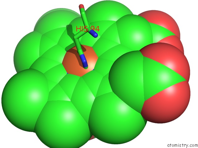
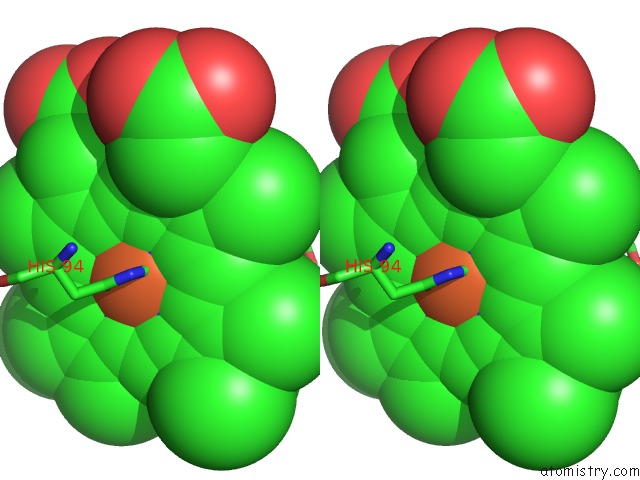
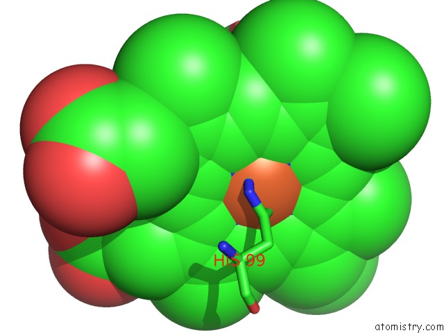
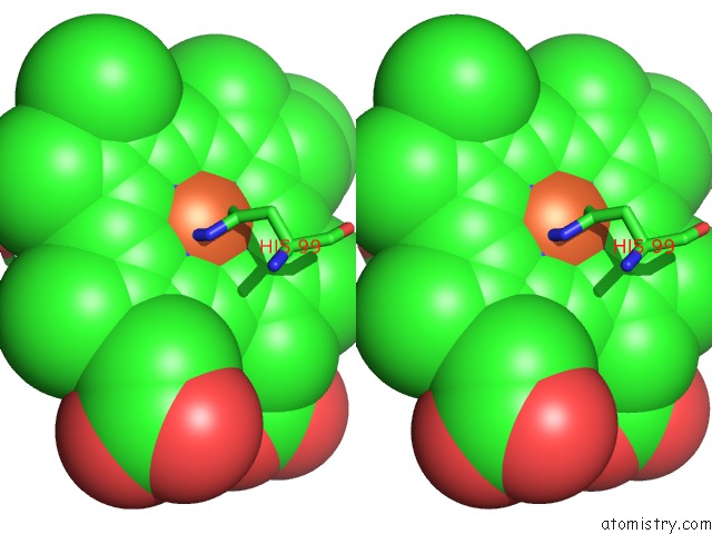
Iron binding site 1 out of 8 in 3wcv
Go back to
Iron binding site 1 out
of 8 in the The Structure of A Deoxygenated 400 kDa Hemoglobin Provides A More Accurate Description of the Cooperative Mechanism of Giant Hemoglobins: Ca Bound Form
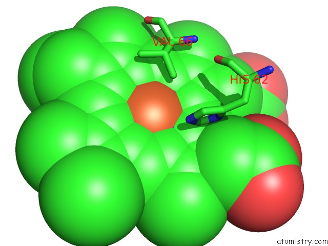
Mono view
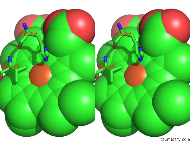
Stereo pair view
Mono view
Stereo pair view
A full contact list of Iron with other atoms in the Fe binding
site number 1 of The Structure of A Deoxygenated 400 kDa Hemoglobin Provides A More Accurate Description of the Cooperative Mechanism of Giant Hemoglobins: Ca Bound Form within 5.0Å range:
|
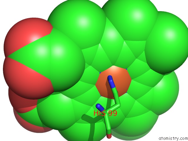
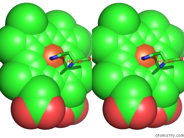
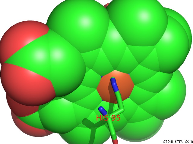
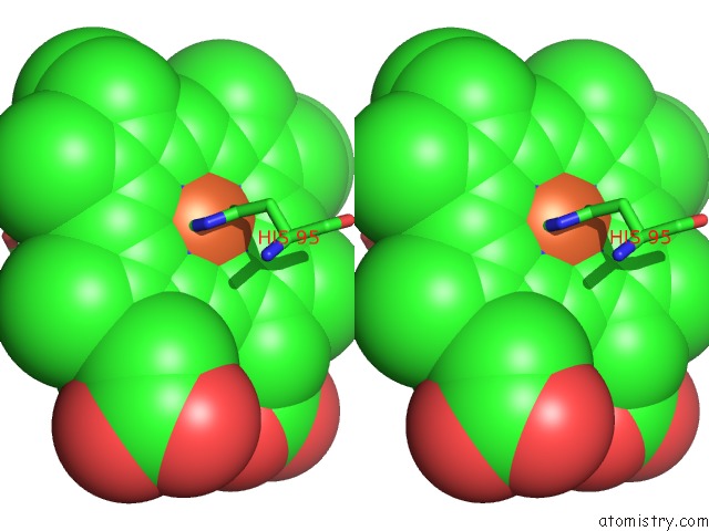
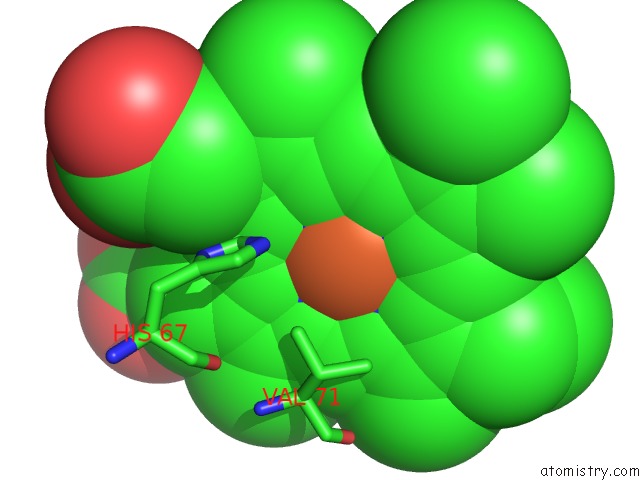
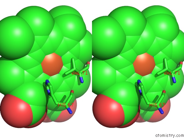
Iron binding site 2 out of 8 in 3wcv
Go back to
Iron binding site 2 out
of 8 in the The Structure of A Deoxygenated 400 kDa Hemoglobin Provides A More Accurate Description of the Cooperative Mechanism of Giant Hemoglobins: Ca Bound Form
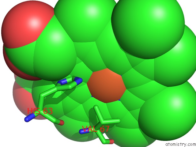
Mono view
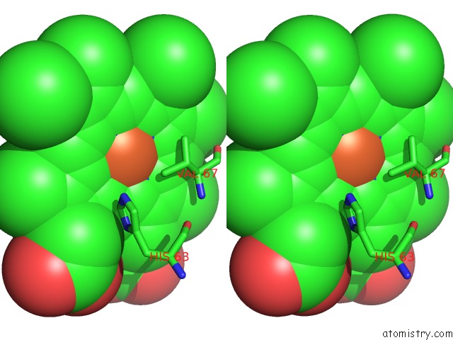
Stereo pair view
Mono view
Stereo pair view
A full contact list of Iron with other atoms in the Fe binding
site number 2 of The Structure of A Deoxygenated 400 kDa Hemoglobin Provides A More Accurate Description of the Cooperative Mechanism of Giant Hemoglobins: Ca Bound Form within 5.0Å range:
|
Iron binding site 3 out of 8 in 3wcv
Go back to
Iron binding site 3 out
of 8 in the The Structure of A Deoxygenated 400 kDa Hemoglobin Provides A More Accurate Description of the Cooperative Mechanism of Giant Hemoglobins: Ca Bound Form
Mono view
Stereo pair view
Mono view
Stereo pair view
A full contact list of Iron with other atoms in the Fe binding
site number 3 of The Structure of A Deoxygenated 400 kDa Hemoglobin Provides A More Accurate Description of the Cooperative Mechanism of Giant Hemoglobins: Ca Bound Form within 5.0Å range:
|
Iron binding site 4 out of 8 in 3wcv
Go back to
Iron binding site 4 out
of 8 in the The Structure of A Deoxygenated 400 kDa Hemoglobin Provides A More Accurate Description of the Cooperative Mechanism of Giant Hemoglobins: Ca Bound Form
Mono view
Stereo pair view
Mono view
Stereo pair view
A full contact list of Iron with other atoms in the Fe binding
site number 4 of The Structure of A Deoxygenated 400 kDa Hemoglobin Provides A More Accurate Description of the Cooperative Mechanism of Giant Hemoglobins: Ca Bound Form within 5.0Å range:
|
Iron binding site 5 out of 8 in 3wcv
Go back to
Iron binding site 5 out
of 8 in the The Structure of A Deoxygenated 400 kDa Hemoglobin Provides A More Accurate Description of the Cooperative Mechanism of Giant Hemoglobins: Ca Bound Form
Mono view
Stereo pair view
Mono view
Stereo pair view
A full contact list of Iron with other atoms in the Fe binding
site number 5 of The Structure of A Deoxygenated 400 kDa Hemoglobin Provides A More Accurate Description of the Cooperative Mechanism of Giant Hemoglobins: Ca Bound Form within 5.0Å range:
|
Iron binding site 6 out of 8 in 3wcv
Go back to
Iron binding site 6 out
of 8 in the The Structure of A Deoxygenated 400 kDa Hemoglobin Provides A More Accurate Description of the Cooperative Mechanism of Giant Hemoglobins: Ca Bound Form
Mono view
Stereo pair view
Mono view
Stereo pair view
A full contact list of Iron with other atoms in the Fe binding
site number 6 of The Structure of A Deoxygenated 400 kDa Hemoglobin Provides A More Accurate Description of the Cooperative Mechanism of Giant Hemoglobins: Ca Bound Form within 5.0Å range:
|
Iron binding site 7 out of 8 in 3wcv
Go back to
Iron binding site 7 out
of 8 in the The Structure of A Deoxygenated 400 kDa Hemoglobin Provides A More Accurate Description of the Cooperative Mechanism of Giant Hemoglobins: Ca Bound Form
Mono view
Stereo pair view
Mono view
Stereo pair view
A full contact list of Iron with other atoms in the Fe binding
site number 7 of The Structure of A Deoxygenated 400 kDa Hemoglobin Provides A More Accurate Description of the Cooperative Mechanism of Giant Hemoglobins: Ca Bound Form within 5.0Å range:
|
Iron binding site 8 out of 8 in 3wcv
Go back to
Iron binding site 8 out
of 8 in the The Structure of A Deoxygenated 400 kDa Hemoglobin Provides A More Accurate Description of the Cooperative Mechanism of Giant Hemoglobins: Ca Bound Form
Mono view
Stereo pair view
Mono view
Stereo pair view
A full contact list of Iron with other atoms in the Fe binding
site number 8 of The Structure of A Deoxygenated 400 kDa Hemoglobin Provides A More Accurate Description of the Cooperative Mechanism of Giant Hemoglobins: Ca Bound Form within 5.0Å range:
|
Reference:
N.Numoto,
T.Nakagawa,
R.Ohara,
T.Hasegawa,
A.Kita,
T.Yoshida,
T.Maruyama,
K.Imai,
Y.Fukumori,
K.Miki.
The Structure of A Deoxygenated 400 kDa Haemoglobin Reveals Ternary- and Quaternary-Structural Changes of Giant Haemoglobins Acta Crystallogr.,Sect.D V. 70 1823 2014.
ISSN: ISSN 0907-4449
PubMed: 25004960
DOI: 10.1107/S1399004714008475
Page generated: Tue Aug 5 08:20:08 2025
ISSN: ISSN 0907-4449
PubMed: 25004960
DOI: 10.1107/S1399004714008475
Last articles
Mg in 4JI2Mg in 4JI3
Mg in 4JHD
Mg in 4JH6
Mg in 4JH8
Mg in 4JH7
Mg in 4JH3
Mg in 4JH5
Mg in 4JF2
Mg in 4JH2